Download presentation
Presentation is loading. Please wait.

1
Terminology, Images & Stuff
FRACTURES Terminology, Images & Stuff Jeannean Rollins, MRC, BSRT, (R)(CV) Associate Professor, Medical Imaging & Radiation Sciences Arkansas State University Jonesboro, AR
(CV) Associate Professor, Medical Imaging & Radiation Sciences. Arkansas State University. Jonesboro, AR.")
2
Objectives Define fracture
Define the 5 descriptors used to classify fractures in long bones Discuss the fractures with “special” names, i.e., eponyms Review the classifications for fractures of the vertebral column Review sample images of fractures

3
Why do we have to study pathology?
We not even allowed to mention diagnosis on images!! An understanding of pathology makes us better care providers and more productive contributors to the healthcare team.

4
Fracture Definition “A disruption of bone caused by mechanical forces applied either directly to the bone or transmitted along the shaft of a bone.” (Eisenberg 131) Eisenberg, R. & Johnson, N. Comprehensive Radiographic Pathology, 5th Edition. Elsevier, St. Louis
 Eisenberg, R. & Johnson, N. Comprehensive Radiographic Pathology, 5th Edition. Elsevier, St. Louis")
5
Radiographic Manifestations
Radiolucent line crossing the bone & interrupting cortical margins Radiopaque line or area due to overlapping bone fragments

6
Secondary Signs of Fracture
Joint effusion Soft tissue swelling Interruption of normal pattern of bony trabeculae

7
“If you can move it, it isn’t broken”

8
“If you can move it, it isn’t broken”

9
The presence or absence of pain &/or the ability to move the part are NOT signs of an underlying fracture Fracture Factoid:

10
Descriptions/Classifications of Fractures
Extent of fracture Direction of fracture line(s) Position of fracture fragments Number of fracture lines Integrity of overlying skin
 Position of fracture fragments. Number of fracture lines. Integrity of overlying skin.")
11
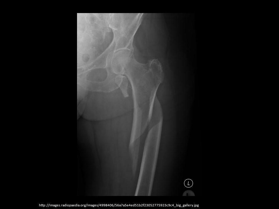
Extent of Fracture Complete Incomplete
results in the discontinuity between 2 or more fragments Incomplete causes only partial discontinuity between fragments, leaving part of cortex in place

12
http://images. radiopaedia

13
http://images. radiopaedia
14
http://images. radiopaedia
15
Direction of Fracture Line(s)
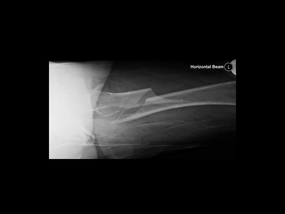
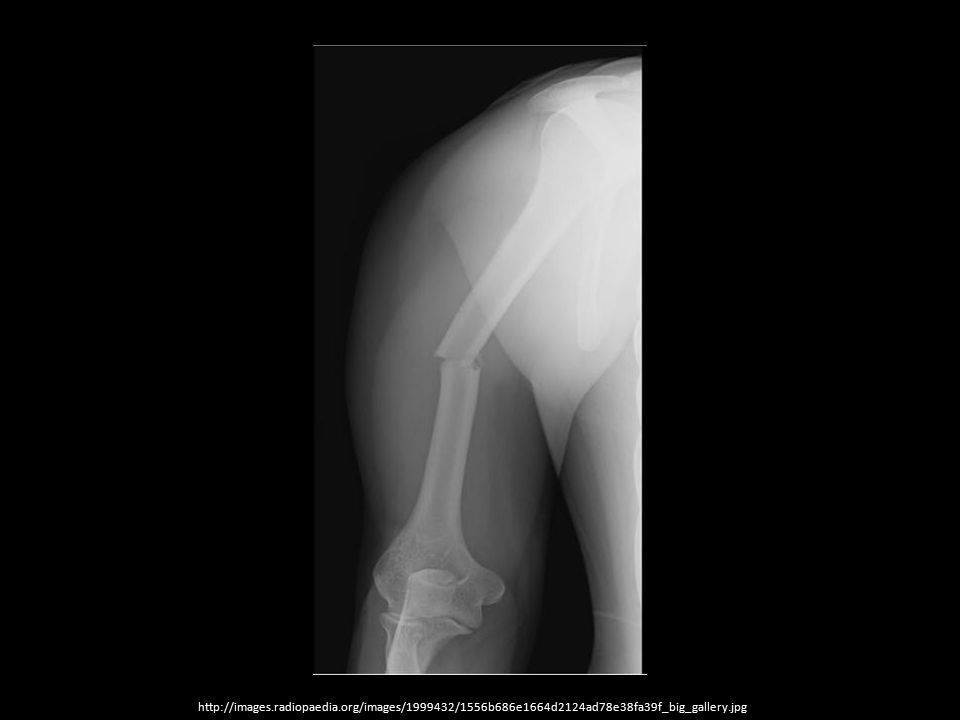
Transverse Runs at right angle to long axis; Usually results from direct blow or pathology Oblique Runs about 45 degrees to long axis; Results from angulation force Spiral Encircles shaft; Caused by torsional force

16
http://images. radiopaedia
18
http://images. radiopaedia
19
http://images. radiopaedia
20
http://images. radiopaedia
21
Position of Fracture Fragments
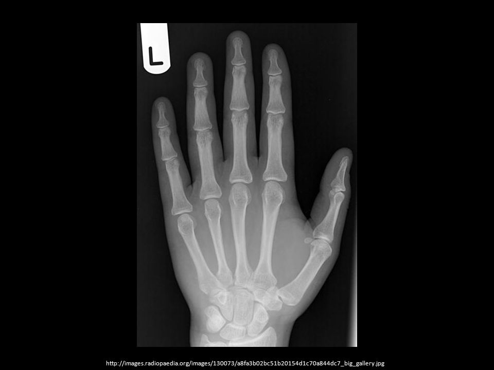
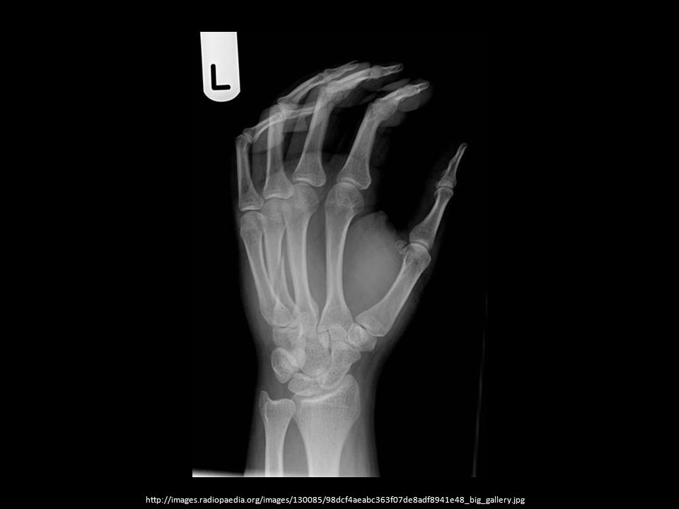
Undisplaced No angulation or separation of fragments Displaced Bone fragments separated; Described in relation of distal fragment in relation to proximal Angulation Indicates angular deformity between axes of major fragments

22
http://images. radiopaedia
23
http://images. radiopaedia
25
http://images. radiopaedia
26
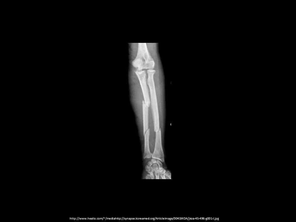
Number of Fracture Lines
Comminuted Describes when there are 2 or more fracture fragments Segmental Consists of a segment of the shaft separated by proximal and distal fracture lines

28
http://www. healio. com/~/mediahttp://synapse. koreamed
29
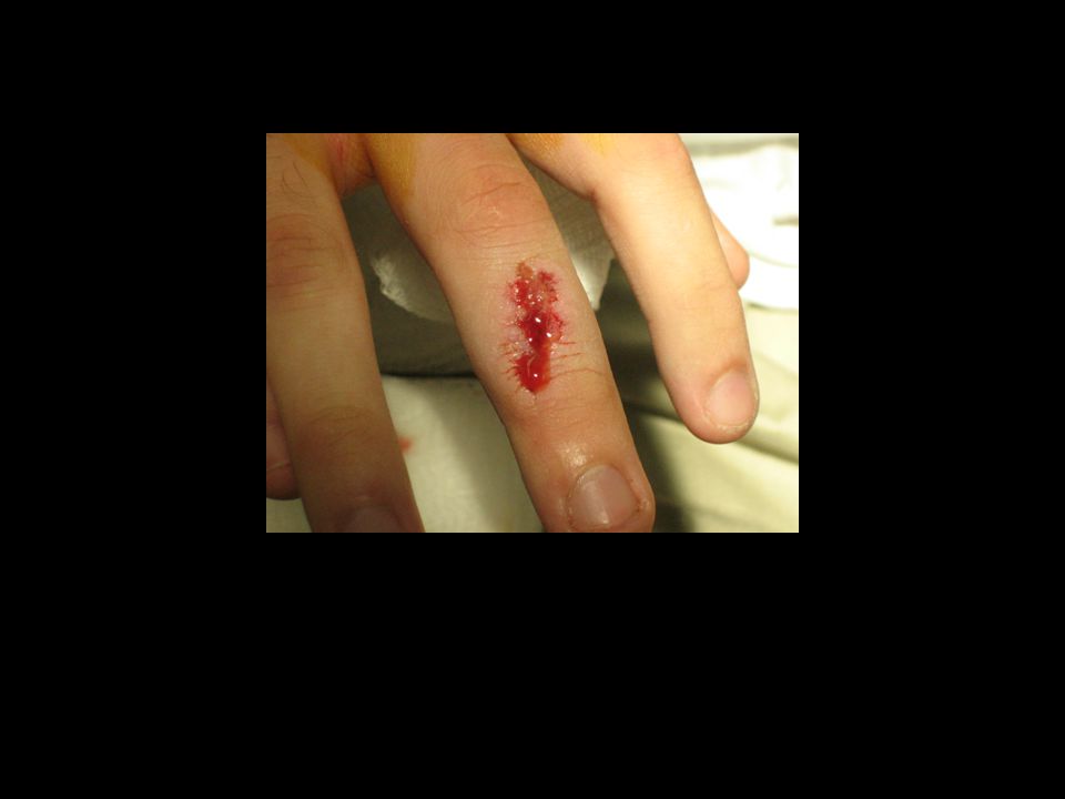
Integrity of Overlying Skin
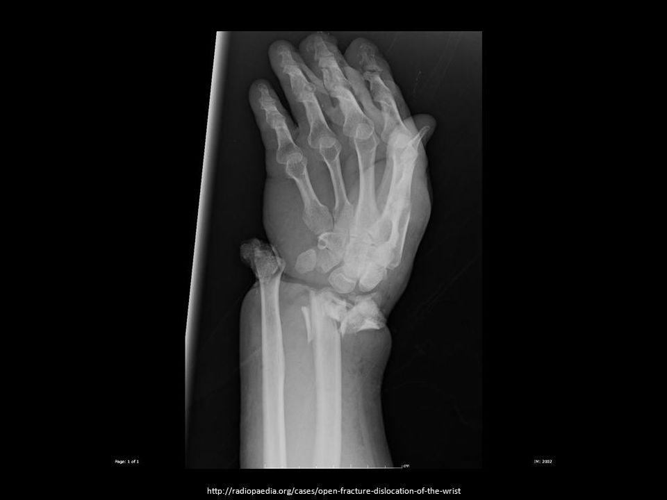
Closed describes when the skin is intact Open/Compound describes when the skin is disrupted; any type of wound over a fracture site, whether or not bone is protruding

32
What precautions do we take during imaging of open fractures?

33
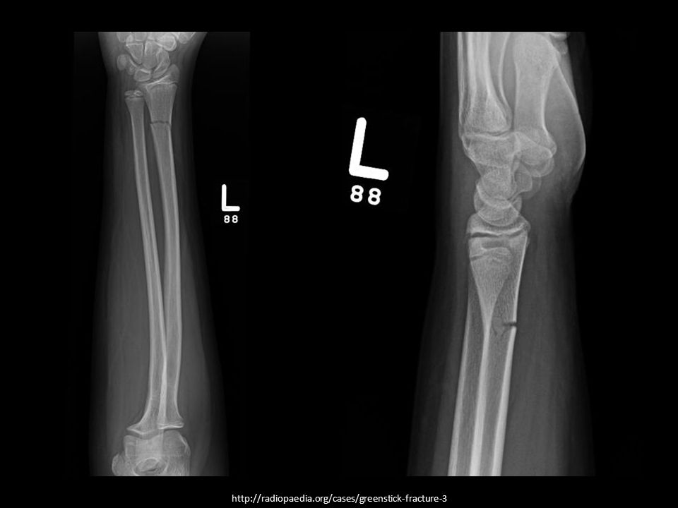
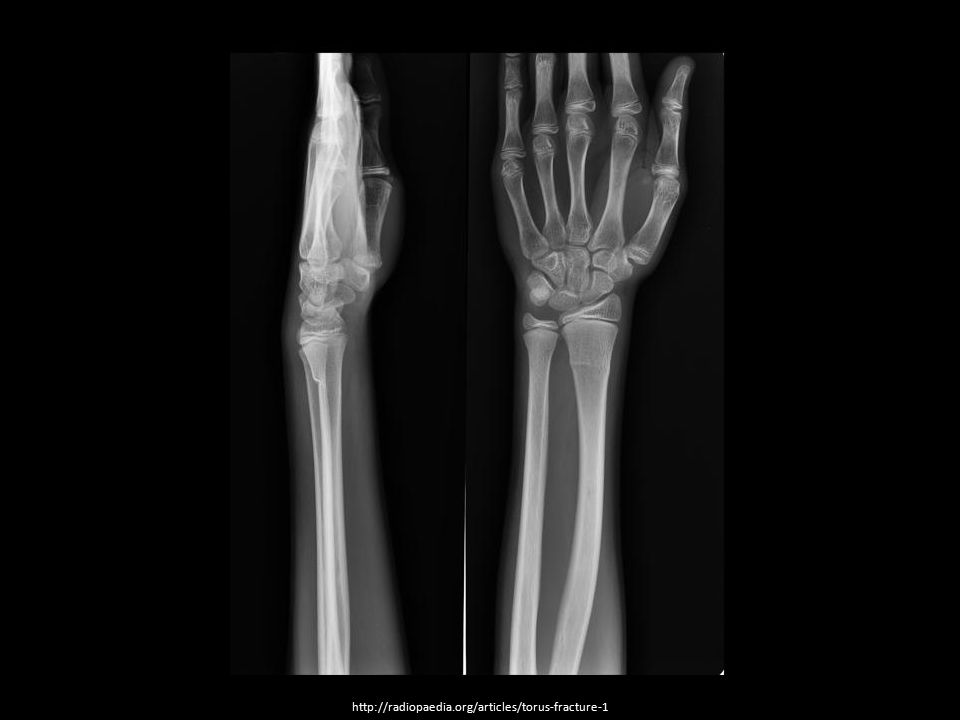
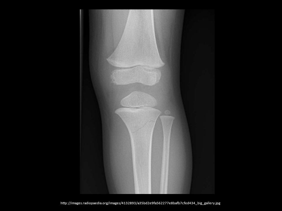
Pediatric Fractures Greenstick Torus/Buckle Salter-Harris
Abbreviated SH Initials followed by a number (I-V) indicating severity I – least severe; V – most severe
 indicating severity. I – least severe; V – most severe.")
36
Salter-Harris fx Involves epiphyseal (growth) plate Greatest concern: Death of the growth plate Causes limb length discrepancy

37
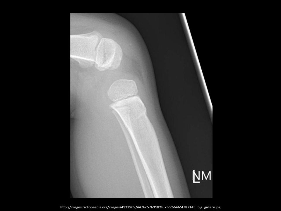
SH-II

38
Upper Limb Common Eponymous Fractures
Boxer Bennett Colles Monteggia Galeazzi Hill-Sachs Bankart

39
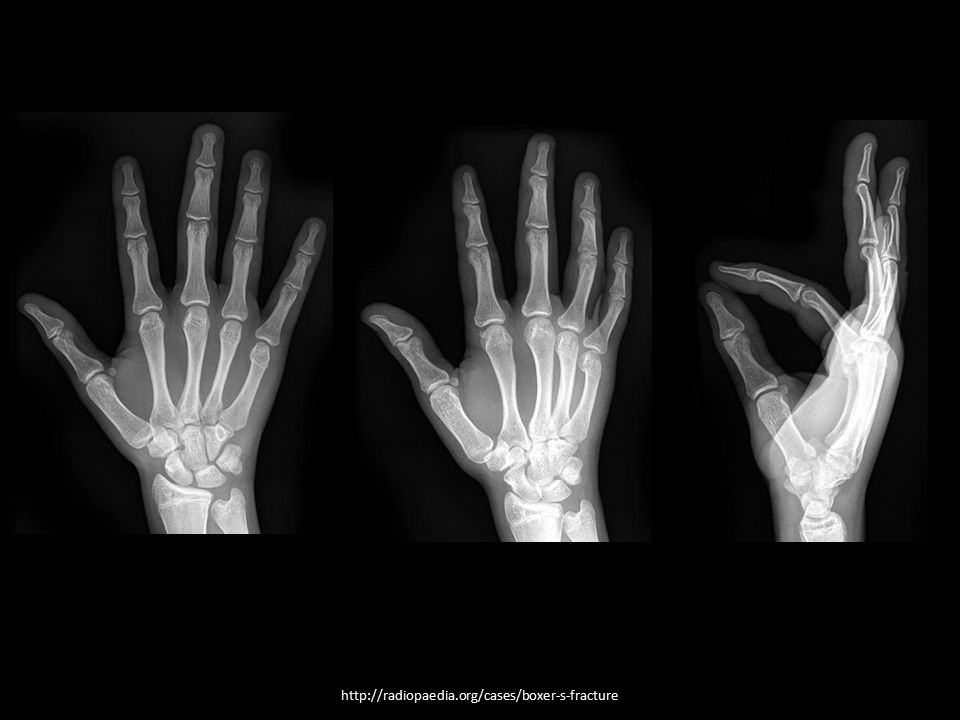
Boxer Fracture Fx of 5th metacarpal w/ palmar (volar) angulation
Name reflects the mechanism of injury Commonly caused by hitting a solid object with a closed fist

41
Bennett Fracture Defined as a fracture at the base of the 1st metacarpal that extends into the CMC joint Also called an intraarticular fracture or a fracture/dislocation Mechanism of Injury: Axial load on a partially flexed thumb

42
Bennett Fracture Critical because incorrect or delayed diagnosis can result in: Early arthritis and pain Loss of some thumb mobility

43
Bennett’s Fracture

44
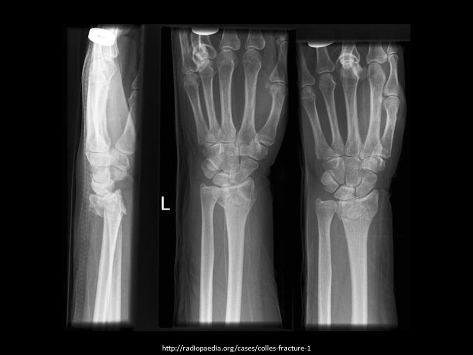
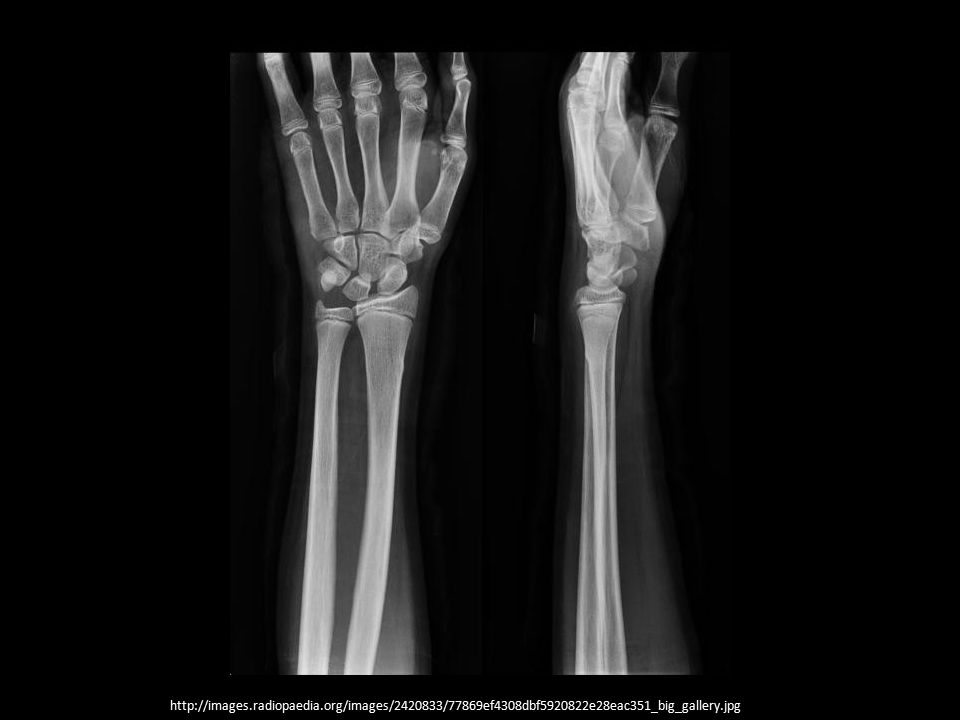
Colles Fracture Most common fracture of the distal radius
Osteoporosis is a risk factor Usually results from a fall on an outstretched hand Dorsal displacement of the distal fragment is characteristic Smith fracture (reverse Colles) has volar displacement
 has volar displacement.")
46
Monteggia/Galeazzi Fractures
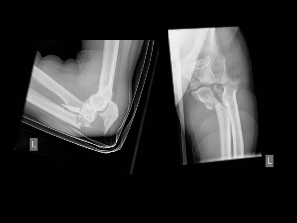
Both are fracture/dislocation injuries of the forearm Monteggia Fx of ulna with dislocation of the radial head Galeazzi Fracture of radius with dislocation of ulnar head

47
Monteggia

48
Galeazzi

49
Hill-Sachs & Bankart Caused by frequent anterior shoulder dislocations
Often occur simultaneously Often requires CT or MRI to diagnose Hill-Sachs Posterorlateral humeral head compression fracture Bankart Fx of inferior glenoid

50
Bankart

51
Hill-Sachs Hill-Sachs

52
Lower Limb Common Fractures
Jones Charcot joint Maisonneuve

53
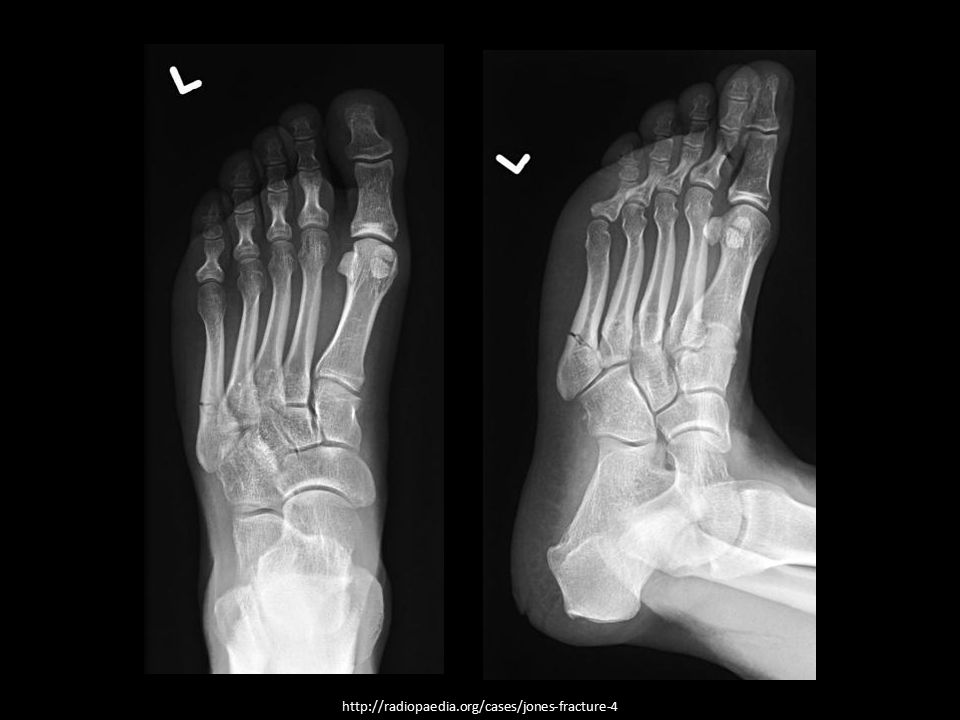
Jones Fracture A transverse fracture at the base of the fifth metatarsal, 1.5 to 3 cm distal to the tuberosity at the metadiaphyseal junction Other common fractures at this site: Stress Avulsion

54
Jones Fracture

56
Charcot Joint AKA: Charcot (Charcot’s) foot, neurotrophic joint, neuropathic joint Progressive degenerative/destructive joint disorder in patients with abnormal pain sensation and proprioception 1 Diabetes is the most common cause in western societies 1- Dähnert W. Radiology review manual. Lippincott Williams & Wilkins. (2007) ISBN:
 ISBN:")
57
Charcot Joint Other causes: syphilis, steroid use, syringomyelia, spinal cord injury, spina bifida, scleroderma, leprosy Radiographic features = 6 D’s Dense bones (sclerosis) Degeneration Destruction (articular cartilage) Deformity metatarsal heads) Debris (loose bodies) Dislocation
 Degeneration. Destruction (articular cartilage) Deformity metatarsal heads) Debris (loose bodies) Dislocation.")
58
46 y/o male. Peripheral neuropathy in type I diabetes mellitus
46 y/o male. Peripheral neuropathy in type I diabetes mellitus. Foot deformity and gait disturbance with minor pain.

59
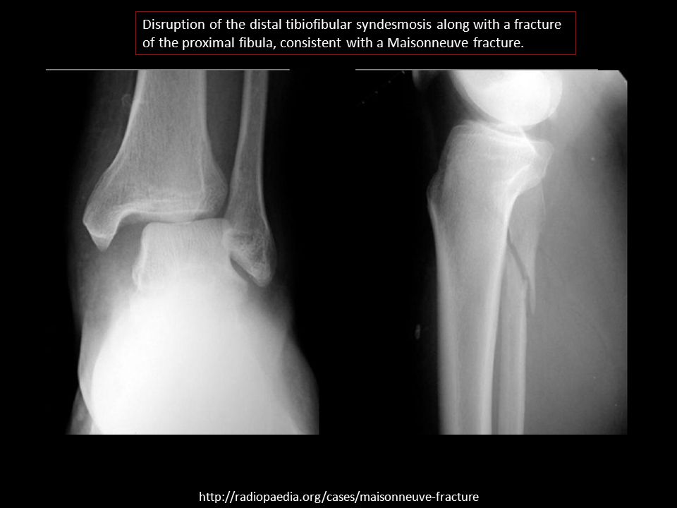
Maisonneuve An unstable fracture typically involving the medial tibial malleolus and/or disruption of the distal tibiofibular syndesmosis along with a fracture of the proximal fibula shaft. The deltoid ligament can be frequently disrupted.

60
Disruption of the distal tibiofibular syndesmosis along with a fracture of the proximal fibula, consistent with a Maisonneuve fracture.
61
Undisplaced spiral fracture through the proximal fibula
Undisplaced spiral fracture through the proximal fibula. Undisplaced transverse fracture through the medial malleolus. Distal tibiotalar joint appears intact.

62
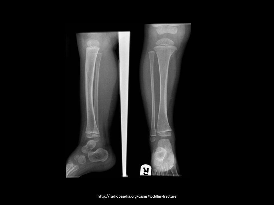
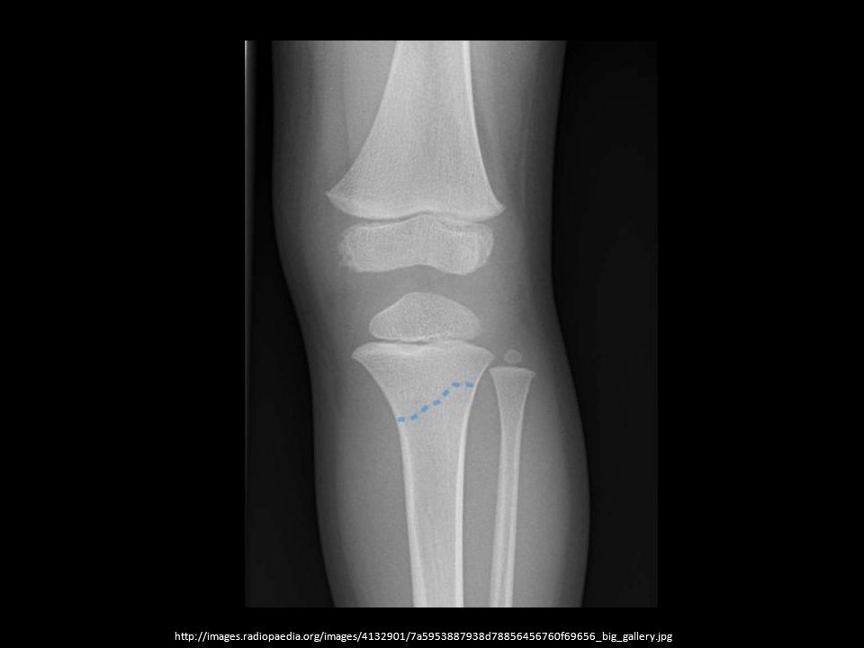
Toddler Fracture Minimally or undisplaced spiral fracture of the tibia
Thought to occur due to new stresses on the bone due to recent ambulation NOT suspicious of child abuse when present in isolation and in the correct age group (9 mos. – 3 yrs.)
")
64
Classifications and Common Types
Spine Fractures

65
Classification of Spine Fractures
Mechanism of Injury Hyperflexion Hyperextension Axial compression Lateral compression Complex injuries 4 Line Method Three Column (Denis) All of these determine stability of spine fracture
 All of these determine stability of spine fracture.")
66
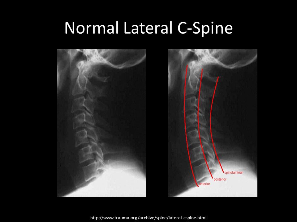
4 Line Method Lines A, B and C should have a smooth curve with no steps or discontinuities. Rotation may cause greater malalignment Line B as compared to Line A > 3.5mm translation anywhere is significant Spinal canal (SC) diameter should be 18mm or greater. Stenosis 14mm or less.
 diameter should be 18mm or greater. Stenosis 14mm or less.")
67
Normal Lateral C-Spine
68
C-Spine Injury

69
Three-Column (Denis) Devised for classification of thoracolumbar fractures Vertebral column divided into three parts based on biomechanical studies related to stability post-traumatic injury

70
Three-Column (Denis) Anterior column Anterior longitudinal ligament
Anterior two-thirds of the vertebral body/intervertebral disc Middle column Posterior one-third of the vertebral body/intervertebral disc Posterior longitudinal ligament Posterior column Facet joints and articular processes Ligamentum flavum Neural arch and interconnecting ligaments Instability - injures two contiguous columns

72
Spine Fractures Cervical Odontoid Thoracolumbar Jefferson Hangman
Clay-shoveler Flexion-teardrop Burst (compression) Odontoid Type I Type II Type III Thoracolumbar Chance
 Odontoid. Type I. Type II. Type III. Thoracolumbar. Chance.")
73
Jefferson Fracture C1 burst fracture
Typical cause – axial load (diving into shallow water) Stable, non-neurologic injury if ligaments are intact AP open- mouth Asymmetry of lateral masses CT &/or MR often needed
 Stable, non-neurologic injury if ligaments are intact. AP open- mouth. Asymmetry of lateral masses. CT &/or MR often needed.")
74
Hangman Fracture Bilateral lamina and pedicle fracture at C2
Usually associated with anterolisthesis of C2 on C3 Most common cause - MVA Lateral c-spine demo’s CT &/or MR often needed

75
Clay-shoveler Fracture
A fracture of the spinous process of a lower cervical vertebra (most commonly, C7) Usually a stress fracture, but acute causes are: Direct force MVA
 Usually a stress fracture, but acute causes are: Direct force. MVA.")
76
Flexion-Teardrop Fracture
Most severe fracture of the c-spine, often causing anterior cervical cord syndrome and quadriplegia Causes: Diving MVA deceleration CT &/or MR required

77
Odontoid Fracture Type I: fracture of the upper part of the dens; rare and potentially unstable Type II: fracture at the base; unstable, and has a high risk of non-union; most common Type III: through the odontoid and into the lateral masses of C2; best prognosis for healing because of the larger surface area of the fracture ~20% of c-spine fractures

78
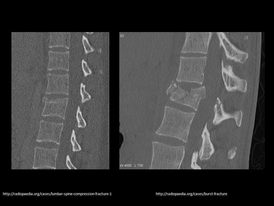
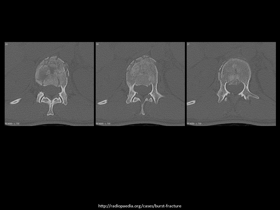
Burst (Compression) Fracture
A type of compression fracture The posterior vertebral body cortex is disrupted and is pushed backward into the spinal canal In the T/L region, tends to occur between T9 and L5 levels Burst fractures may be stable or unstable

80
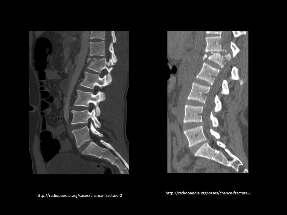
Chance Fracture Bony injuries that extend all the way through the spinal column The most common history is a MVA or fall from a height Back seat passenger w/ a lap seatbelt The middle and posterior columns are typically disrupted High incidence of associated intra-abdominal injuries

82
Questions? Comments? jrollins@astate.edu

Similar presentations