Download presentation
Presentation is loading. Please wait.

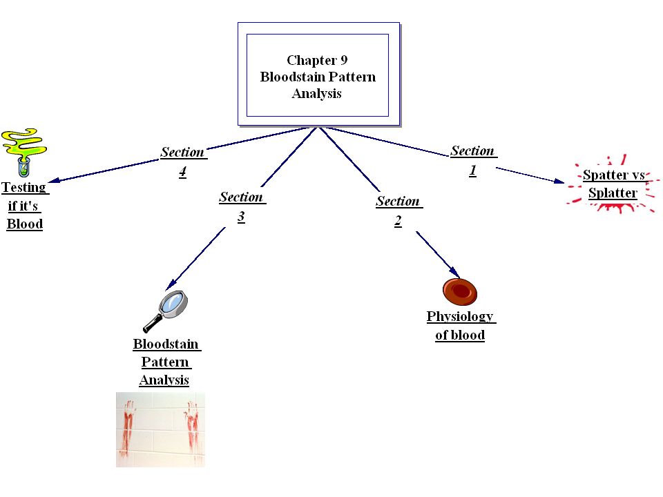
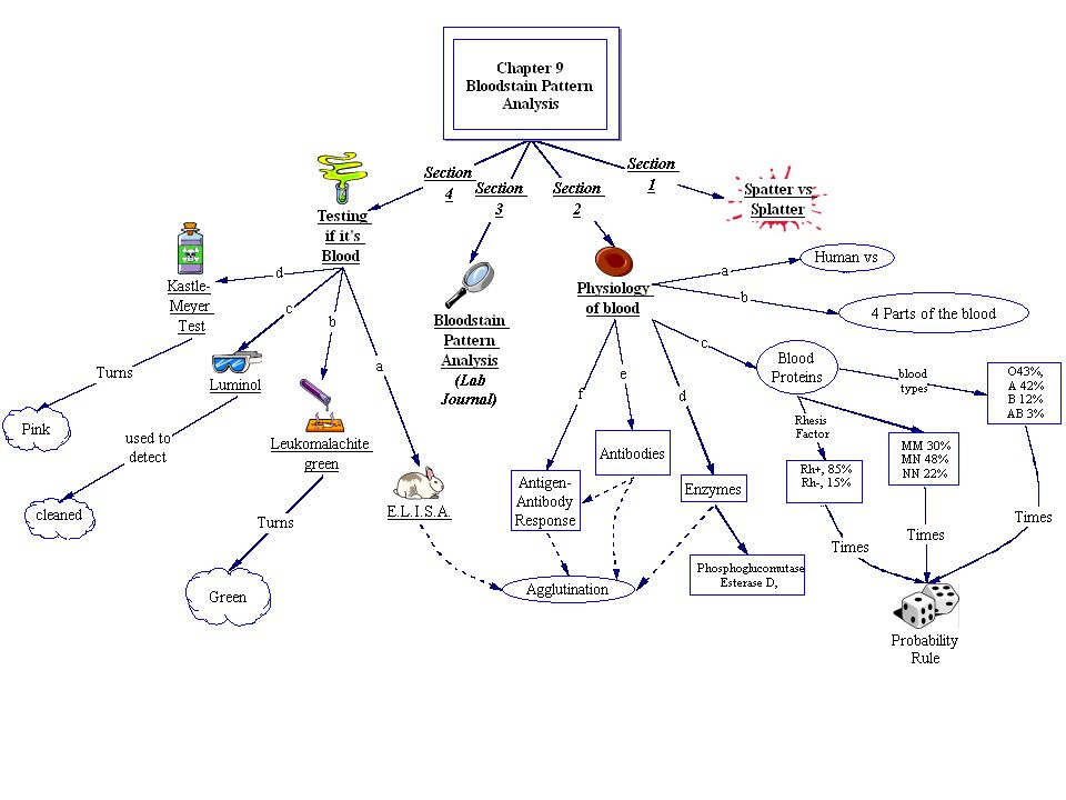
2
Spatter vs splatter Splatter: random, unorganized
Spatter: Not random, affect by gravity, predictable Blood stain Pattern: The pattern of a stain and the quantity of blood present can be important clues to the nature of the accident or crime. BSP Interpretation: what does the blod tell you?

3
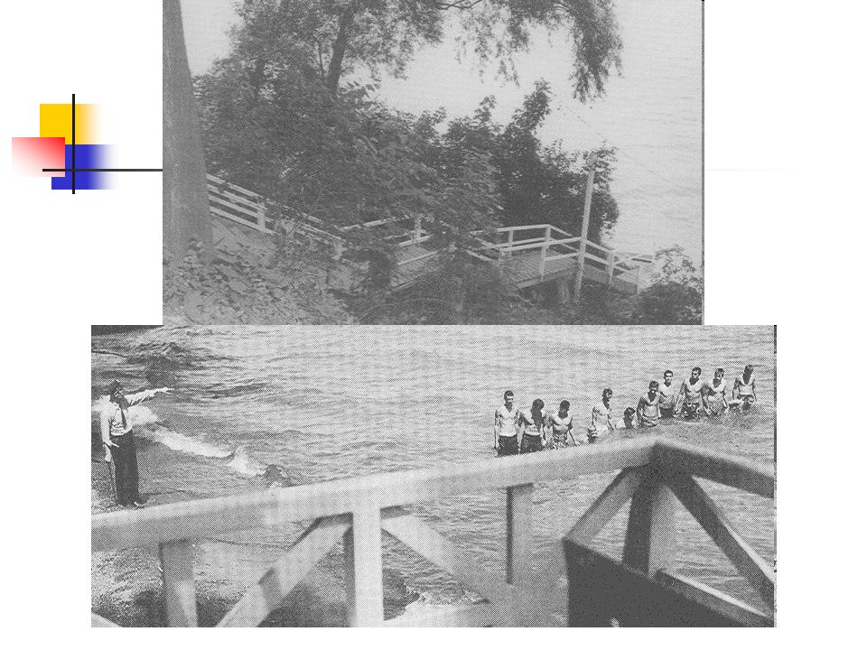
The Sheppard Family Story

4
Dr Sam and Marilyn Sheppard
35 blows, 28 to the head Unknown instrument 12> inches None of these blows were fatal 2 broken teeth Torn fingernail. Blood found on most of the walls along with covering a dresser. Dr. Sam Sheppard: Bruises chipped teeth fractured vertebra in his neck. Found shirtless

5
Crime Scene pictures Spatter ? Sheppard Images

6
Crime Scene pictures

8
Questions What Biological evidence will blood provide,, in the bedroom? What information does the Blood stain evidence provide?

9
Dr Sam Sheppard July 4 1954, Reading:
HW link: main site: Police report:

10
Do Now: What Biological evidence will blood provide,, in the bedroom?
DNA Blood type(s) Tool marks/ voids Toxicological Reports Diseases Spatter marks (Blood trails, movements)
 Tool marks/ voids. Toxicological Reports. Diseases. Spatter marks (Blood trails, movements)")
11
What can Spatter (Bloodstain Evidence) Evidence reveal:
.

12
What can Spatter (Bloodstain Evidence) Evidence reveal:
Origin(s) of bloodstain Position of victim & assailant Movement of victim & assailant Number of blows/shots Distance of bloodstain from target Direction from which blood impacted Speed with which blood left its source
 of bloodstain. Position of victim & assailant. Movement of victim & assailant. Number of blows/shots. Distance of bloodstain from target. Direction from which blood impacted. Speed with which blood left its source.")
14
Serology The study of antigen-antibody reactions.
Tells us human vs. nonhumans Serologists Questions Is the sample blood? Is the sample animal blood? If animal blood, from what species? If human blood, what type? Can the sex, age, and race of the source of blood be determined?

15
History of Blood!!! With early Transfusions =instant death due to Coagulation , Karl Landsteiner introduced the A-B-O system ,Alexander Weiner, Rhesus monkey, Rh Factor was and over 100 factors actually must be considered when performing a transfusion. Most people are only familiar with the Rh factor, which is technically the D antigen. There are more than 256 antigens, and 23 blood group systems based on association with these antigens. Us Populations: O 43%, A 42%, B 12 %, AB 3%

16
Blood Typing

17
Structure of Blood: Plasma, mostly water Cells Erythrocytes: (RBC)
Leukocytes: (WBC) Platelets
 Platelets.")
18
Major components of Blood, Plasma:
Straw colored liquid consisting mainly (90%) of water and (7%) dissolved proteins. Can be found outside of the circulatory system. Also transports: Proteins: (albumin, globulins, fibrinogen) Salts, Glucose Amino acids Fatty acids, Vitamins, Hormones, Cellular wastes
 of water and (7%) dissolved proteins. Can be found outside of the circulatory system. Also transports: Proteins: (albumin, globulins, fibrinogen) Salts, Glucose. Amino acids. Fatty acids, Vitamins, Hormones, Cellular wastes.")
19
DNA in BLOOD DNA can be extracted from blood (if white blood cells which always contain a nucleus are present), and also from sperm, bone marrow, tooth pulp, and hair roots.
, and also from sperm, bone marrow, tooth pulp, and hair roots.")
20
Blood, however, is commonly used in DNA testing, as per the following steps:
Blood samples are collected from the victim, defendant, and crime scene White blood cells are separated from red blood cells DNA is extracted from the nuclei of white blood cells A restrictive enzyme is used to cut fragments of the DNA strand DNA fragments are put into a bed of gel with electrodes at either end Electric current sorts DNA fragments by length An absorbent blotter soaks up the imprint; it is radioactively treated, and an X-ray photograph (called an autoradiograph) is produced
 is produced")
21
Liquid Blood Physical properties Behaves as a projectile in motion 2
viscosity surface tension specific gravity Behaves as a projectile in motion biology, physics, maths

22
Surface Tension Resistance to penetration & separation
3 Surface Tension Resistance to penetration & separation Surface acts to reduce surface area Smallest SA to Volume ratio is offered by sphere

23
Dripping Blood 4 Blood trickles downwards
Blood drop grows until Wt (G) > S.T. Single drop breaks free (teardrop shape) Surface tension pulls in vertically And horizontally Shape settles into sphere (0.05 ml) Does not break up until impact
 > S.T. Single drop breaks free (teardrop shape) Surface tension pulls in vertically. And horizontally. Shape settles into sphere (0.05 ml) Does not break up until impact.")
24
. . . Drop size 5 Standard drop size 50ul (0.05ml)
Rapid bleeding gives slightly larger drop Shaking/movement casts off smaller drops . . .

25
Terminal Velocity v Distance Fallen (metric)
6 Terminal Velocity v Distance Fallen (metric)
")
26
Terminal Velocity v Distance Fallen (imperial)
7 Terminal Velocity v Distance Fallen (imperial)
")
27
Free Falling Blood Droplets
8 Free Falling Blood Droplets 0.06 ul 1.1 mm 0.5 to 0.65 m . 2.2 m/s 0.12 ul 1.32 mm 0.84 to 1 m . 3.3 m/s 0.5 ul 2.12 mm 2.4 to 3 m . 4.6 m/s . 50 ul 4.6 mm 7.5 m/s 4.2 to 5.4 m

28
Shape & Size of Bloodspot
9 Shape & Size of Bloodspot Depends mostly on nature of target surface texture (rough or smooth) porous or non porous Size is related to distance fallen, provided: standard 50 ul drop of blood There is little change in spot diameter beyond a fall distance of 1.2 m
 porous or non porous. Size is related to distance fallen, provided: standard 50 ul drop of blood. There is little change in spot diameter beyond a fall distance of 1.2 m.")
29
Shape & Size of Bloodspot
9 Shape & Size of Bloodspot

30
Hat information does this questionable drop pelt provide?
9 Hat information does this questionable drop pelt provide?

31
Height Fallen 10 Single drops of blood falling from fingertip onto smooth cardboard from various heights. No change in diameter beyond 7 ft. Adapted from Introduction to Forensic Sciences, W. Eckert, CRC, 1997

32
Effect of Target Surface
11 Effect of Target Surface . . . Spreads out smoothly ST of spreading edge is broken by irregular surface

33
Experiments with Falling Blood Droplets
12 blood dropper ruler Height Target Surface Fabric (theatre green) rough paper towel paper whiteboard Terazzo floor
 rough paper towel. paper. whiteboard. Terazzo floor.")
34
Single drop of blood falling from various heights (m) onto various surfaces
13 0.5 1 2 3 0.5 1 2 3 Height/Surface smooth floor paper towel fabric

35
IMPACT ANGLE DETERMINATION
ANGLE of IMPACT is the acute angle formed between the direction of the blood drop and the plane of the surface it strikes

36
Angle of Impact 14 Adapted from Introduction to Forensic Sciences,
W. Eckert, CRC, 1997

37
Angle of Impact 14 Gravitational dense zone at lower edge 90 80 70
60 50 40 Gravitational dense zone at lower edge 20 30 10 Adapted from Introduction to Forensic Sciences, W. Eckert, CRC, 1997

38
. Wave Cast-off 15 Tail of elongated stain
points in direction of travel . Tail of wave cast-off points back to parent drop Parent drop wave cast-off

39
Point of Convergence 16

40
Point of Convergence 16

41
Point of Convergence 17 5 ml blood squirted from a
syringe from height of 1 m Point of Convergence

42
Point of Origin 1 18 Distance from point of convergence
Height above point of convergence Origin length width Angle of impact = arc sin W/L 85 60 45 30

43
Tracing Origin of Bloodspots
19 Tracing Origin of Bloodspots Point of convergence method 2 dimensional image Point of origin method adds 3rd dimension to image In practice: use of string & protractor at scene use of computer at laboratory

44
Blood Spatter Low velocity (5 f/s, 1.5 m/s)
20 Blood Spatter Low velocity (5 f/s, 1.5 m/s) e.g. free-falling drops, cast off from weapon Medium velocity ( f/s, m/s) e.g. baseball bat blows High velocity (>100 f/s, 30 m/s) e.g. gunshot, machinery
 e.g. free-falling drops, cast off from weapon. Medium velocity ( f/s, m/s) e.g. baseball bat blows. High velocity (>100 f/s, 30 m/s) e.g. gunshot, machinery.")
45
Herbert Leon MacDonell,
Laboratory of Forensic Science, P.O. Box 1111, Corning, New York, 14830, USA 21

46
Low Velocity Blood Spatter
22 Low Velocity Blood Spatter Blood source subjected to LV impact < 5 f/s (1.5 m/s) Spot diameter: mostly mm some smaller, some larger Free-falling drops (gravity only) Cast off from fist, shoe, weapon Dripping Splashing Arterial spurting
 Spot diameter: mostly mm. some smaller, some larger. Free-falling drops (gravity only) Cast off from fist, shoe, weapon. Dripping. Splashing. Arterial spurting.")
47
Cast-off from Weapon First blow causes bleeding
23 Cast-off from Weapon First blow causes bleeding Subsequent blows contaminate weapon with blood Blood is cast-off tangientially to arc of upswing or backswing Pattern & intensity depends on: type of weapon amount of blood adhering to weapon length of arc

48
24 Downswing of Hammer

49
25 Cast-off from Weapon ceiling

50
Overhead swing with bloodied metal bar
26 Overhead swing with bloodied metal bar

51
27 Cast-off Pattern (1/2)
")
52
28 Cast off Pattern (2/2) 1 2 3
 1 2 3")
53
29 What does this tell you?

54
Cast off Pattern (2/2) ? Sequence
29 Cast off Pattern (2/2) ? Sequence
 Sequence.")
55
Cast off Pattern (2/2) ? Sequence
30 Cast off Pattern (2/2) ? Sequence 1 (4 spots) 2 (3 spots) 3 (2 spots) If weapon does not pick up more blood, spatter from subsequent backswings becomes progressively less. In practice weapon picks up more blood with each successful blow.
 Sequence. 1. (4 spots) 2. (3 spots) 3. (2 spots) If weapon does not pick up more blood, spatter from subsequent backswings becomes progressively less. In practice weapon picks up more blood with each successful blow.")
56
Three overhead swings with hatchet
31 Three overhead swings with hatchet

57
Cast-off & medium velocity spatter
32

58
Cast-off & medium velocity spatter 2
33 Cast-off & medium velocity spatter 2

59
Cast-off Pattern ? Object
34 Cast-off Pattern ? Object

60
Cast-off Pattern from Hand
35 Cast-off Pattern from Hand

61
Cast-off pattern from bloodied hand swung in front of target
36 6” ruler

62
Drip Pattern 37 Free-falling drops dripping into wet blood Large irregular central stain Small round & oval satellite stains . . . . . . . . . . .

63
38 Drip 1: Blood dripping into itself from height of 1 m (8 drops)
")
64
Blood dripping into itself from height of 1 m (8 drops)
39

65
40 Dripping onto steps

66
Splash Pattern Volume > 1 ml
41 Volume > 1 ml Subjected to LV impact Thrown Tipped Large central irregular area surrounded by elongated peripheral spatter pattern

67
42 Splash 1 5 ml blood squirted from a syringe from a height of 1 m

68
5 ml blood squirted from a
syringe from a height of 1 m Splash 2 43

69
5 ml blood squirted from a syringe from a height of 1 m
44 Splash 3

70
Splash onto vertical surface
45 Splash onto vertical surface 10 ml blood thrown 1 m onto a vertical target surface 6” ruler

71
46 Stamping in blood 1 Area seen in close-up in next slide

72
Stamping in blood Close-up of heel area
47 Stamping in blood Close-up of heel area

73
48 Blood pool (10 drops) before stamping Stamp 1
 before stamping Stamp 1")
74
49 Blood pool (10 drops) after stamping Stamp 2
 after stamping Stamp 2")
75
Arterial Spurt Pattern
50 Arterial Spurt Pattern Blood exiting body under arterial pressure Large stains with downward flow on vertical surfaces wave-form of pulsatile flow may be apparent

76
51 spatter Small arterial spurt broken pottery

77
Neck incisions (scene)
52

78
Medium Velocity Blood Spatter
54 Medium Velocity Blood Spatter Blood source subjected to MV impact ( f/s, m/s) Spot diameter: mostly mm Blows with weapon (e.g. baseball bat)
 Spot diameter: mostly mm. Blows with weapon (e.g. baseball bat)")
79
Medium velocity blood spatter
Medium velocity blood spatter. Point of impact 15 cm in front of vertical target surface 55 6” ruler

80
Flick 1: Blood flicked between middle finger & thumb
onto a vertical smooth surface from a distance of 15 cm Flick 1: 56

81
Flick 2: Blood flicked between middle finger & thumb
onto a vertical smooth surface from a distance of 15 cm Flick 2: 57

82
High Velocity Blood Spatter
58 High Velocity Blood Spatter Blood source subjected to HV impact > 100 f/s, 30 m/s Fine mist: spot size < 0.1 mm Small mass limits spread to 1 m !Some larger droplets reach further Gunshot back-spatter from entry wound forward spatter from exit wound High speed machinery

83
Gunshot: back& forward spatter
59 Bloodstained foam held just above target surface. Bullet passing L to R just above sheet bullet exits foam Bullet enters foam bullet Back-spatter on entry Forward spatter on exit

84
Gunshot Back Spatter Arises from entrance wound
60 Arises from entrance wound Passes back towards weapon & shooter Seen only at close range of fire Seen on: inside of barrel exterior of weapon hand, arm, chest of shooter

85
Back spatter on steadying hand
61 Back spatter on steadying hand

86
Gunshot Forward Spatter
62 Gunshot Forward Spatter Arises from exit wound Passes forwards in same direction as shot More copious than back-spatter Can be seen at any range of fire Seen on nearby surfaces, objects, persons especially on wall behind victim

87
Forward spatter (5 ms after bullet impacted at 1000 f/s)
63 bullet blood soaked target 2.5 cm

88
Forward spatter onto target placed 15 cm behind point of HV bullet impact (bullet passing towards screen) 1 64 6” ruler

89
Forward spatter (closer view)
65

90
Forward spatter (closest view)
66 5 mm

91
Wipe Patterns Object moves through a wet bloodstain
67 Wipe Patterns Object moves through a wet bloodstain Feathered edge suggests direction

92
Transfer Patterns Wet, bloodied object contacts a secondary surface
68 Wet, bloodied object contacts a secondary surface Transfer from: hand, fingers shoes, weapon hair Transfer to: walls, ceilings clothing, bedding Produces mirror-image of bloodied object

93
Transfer from hair (hair-swipe) 1
69

94
Transfer from hair (hair-swipe) 2
70

95
Flow Patterns Blood flows horizontally & vertically
71 Flow Patterns Blood flows horizontally & vertically Altered by contours, obstacles Often ends in pool

96
Flow pattern 72

97
Bloodspots on trousers
78 Bloodspots on trousers

98
Serology The analysis of the properties and effects of serums (blood, semen, saliva, sweat, or fecal matter) is called serology.
 is called serology.")
99
Immunoassay techniques
Looking for Drugs, toxins, antibodies Antibodies not found in humans are synthesized Usually inject compound with drug (that you are testing for) into an animal Why???? Animal makes antibodies because it is a foreign substance
 into an animal. Why Animal makes antibodies because it is a foreign substance.")
100
Immunoassay techniques
Enzyme Multiplied Immunoassay Technique (EMIT): Detection of drugs through a antigen-antibody reaction. Radioimmunoassay (RIA) uses drugs that are labeled with radioactive drugs SO DETECTION occurs with any other related forms of that drug type!!!
: Detection of drugs through a antigen-antibody reaction. Radioimmunoassay (RIA) uses drugs that are labeled with radioactive drugs. SO DETECTION occurs with any other related forms of that drug type!!!")
101
Immunoassay techniques ~~EMIT screening
Add subjects urine antibodies to detect material. For methadone: add methadone antibodies to the urine. [Conc] of antibodies not used indicates concentration used by the drug providing a related methadone concentration

102
Immunoassay techniques, ~~EMIT screening
Marijuana's major active agent THC Tetrahydrocannabinol (3- 4.5%) Liquid Hashish oil 8-22 % THC Metabolized into THC-9-carboxlic acid Is detectable THC-9 in smokers urine is<1 mg (millionth of a gram) 2-5 days…10 days (sometimes)
 Liquid Hashish oil 8-22 % THC Metabolized into THC-9-carboxlic acid. Is detectable. THC-9 in smokers urine is<1 mg (millionth of a gram) 2-5 days…10 days (sometimes)")
103
Animal Responses: Polyclonal antibodies: antibodies produced by injecting animals with a specific antigen . A series of antibodies. A series of antibodies are produced responding to a variety of different sites on the antigen. Monoclonal Antibodies: collection of identical antibodies that interact with a single antigen n site. Mass produced by HYBIDOMA CELLS: spleen-cancer hybrids made in limitless supply

105
It’s Red so it must be blood, right?
When found at a CS, you must determine: If it is blood? Human vs animal. How closely it can be associated with a specific individual?

106
Crime Scene analysis of blood
Confirming the stain is blood (Presumptive tests) Luminol Kastle Meyer test Leukomalachite green Hemastix ® Confirming the blood is HUMAN ELISA/Precipitin test
 Luminol. Kastle Meyer test. Leukomalachite green. Hemastix ® Confirming the blood is HUMAN. ELISA/Precipitin test.")
107
Presumptive tests: A simple test for a given substance using a reagent that changes color when mixed with the substance under investigation. Presumptive tests are not definitive and further confirmatory tests are always required. They are used extensively in forensic science. In general analytical chemistry, presumptive tests are often called spot tests. The first test is simply the use of a powerful light moved across every surface of a crime scene. That yields possible traces for visual inspection.

108
1. Luminol Luminol Reagent:
Tests for by production of light rather than color. Extremely sensitive and can detect minute amounts of blood DOES NOT interfere with subsequent DNA analysis

109
Characterization of blood stain

110
1. Luminol Reaction RBC contain hemoglobin Mix luminol + Peroxide
The iron in homoglobin acts as a catalyst speeding up the reaction between Peroxide and luminol. As reaction progresses, light is generated for about 30 seconds (room should be dark)
")
111
1. Luminol sprayed across the scene because it reacts to blood by making it luminescent. It only takes about five seconds. The procedure requires that the room be considerably darkened in order to see the faint bluish glow, and the intensity of the glow increases proportionately to the amount of blood present. It works even with old blood or diluted stains, and can illuminate smear marks where blood has been wiped away. However, there is one problem with this test: luminol can destroy the properties of the blood that investigators need for further testing. Its use is limited to proving that blood is present even if not visible.

112
2. The Kastle-Meyer Color Test
uses a solution of phenolphthalein and hydrogen peroxide on a piece of filter paper, and when blood of any quantity is present, it turns pink. However, it also turns pink in the presence of potatoes or horseradish, so care must be taken at the scene.

113
Characterization of blood stain
3. Hemastix ® is a dipstick for blood Moisten with distilled water and dipped into the sample Positive presence of blood detected if stick turns green

114
Characterization of blood stain
5. Precipitin Tests: (10-15 years) Serum for the precipitin test is obtained from rabbits which have produced antibodies to destroy a small quantity of human blood injected into them. A drop of this anti-human serum is added to suspect blood, which will precipitate its protein if it is of human origin. Electrophoretic method: ?? Western blotting test... analysis can detect one protein in a mixture of any number of ... Western blotting tells you how much protein has accumulated in cells
 Serum for the precipitin test is obtained from rabbits which have produced antibodies to destroy a small quantity of human blood injected into them. A drop of this anti-human serum is added to suspect blood, which will precipitate its protein if it is of human origin. Electrophoretic method: Western blotting test... analysis can detect one protein in a mixture of any number of ... Western blotting tells you how much protein has accumulated in cells.")
115
Precipitin Tests: history
Investigators use the precipitin test to determine whether the blood is of animal or human origin. German biologist Paul Uhlenhuth discovered that if he injected protein from a chicken egg into a rabbit, and then mixed serum from the rabbit with egg white, the egg proteins separated from the liquid to form a cloudy substance known as precipitin. In other words, it forms an antibody. In the forensic test for human blood: either a sample of the suspect blood is put into a test tube over the rabbit serum or it's used in the "gel diffusion" test, where it's placed in gel on a glass slide next to a sample of the reagent (anti-human serum). Passing an electric current through the glass, the protein molecules filter into the gelatin and toward each other. A line forms where they meet---called a precipitin line---that means the sample is human blood.
. Passing an electric current through the glass, the protein molecules filter into the gelatin and toward each other. A line forms where they meet---called a precipitin line---that means the sample is human blood.")
116
Precipitin Tests: history
In 1925, another blood-related discovery important to criminal investigation was made. Around 80 percent of the human population were found to be "secretors," which means that the specific types of antigens, proteins, antibodies, and enzyme characteristic of their blood can be found in other bodily fluids and tissues. In the case of a secretor, investigators can tell the blood type by examining the saliva, teardrops, skin tissue, urine, or semen. In a rape case, for example, where the perpetrator is a secretor, potential suspects can be narrowed down through blood type analysis

117
Characterization of blood stain
5. ELISA/Precipitin Test: Human Antiserum determines if blood is from animal or human origin. HOW does it work? Remember ANTIGEN-Antibody Reaction???????

Similar presentations


>")


 of bloodstain Distance of bloodstain from target Direction from which blood impacted Speed with which blood.>")


