Download presentation
Presentation is loading. Please wait.

1
X-RAY DIFFRACTION (XRD)
MMS KARAKTERISASI MATERIAL + LAB X-RAY DIFFRACTION (XRD) Dr. Ir. A. Herman Yuwono, M. Phil. Eng. Departemen Metalurgi dan Material Fakultas Teknik Universitas Indonesia Tel: +(62 21) Fax : +(62 21)
 Dr. Ir. A. Herman Yuwono, M. Phil. Eng. Departemen Metalurgi dan Material Fakultas Teknik Universitas Indonesia. Tel: +(62 21) Fax : +(62 21)")
2
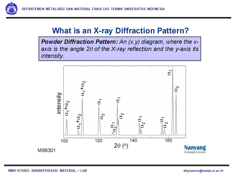
WHAT XRD?

3
Figure caption : X-ray diffraction photograph for a single crystal of magnesium. Schematic diagram illustrating how the spots (i.e., the diffraction pattern) in (a) are produced. The lead screen blocks out all beams generated from the x-ray source, except for a narrow beam traveling in a single direction. This incident beam is diffracted by individual crystallographic planes in the single crystal (having different orientations), which gives rise to the various diffracted beams that impinge on the photographic plate. Intersections of these beams with the plate appear as spots when the film is developed. The large spot in the center of (a) is from the incident beam, which is parallel to a [0001] crystallographic direction. It should be noted that the hexagonal symmetry of magnesium’s hexagonal close-packed crystal structure is indicated by the diffraction spot pattern that was generated.
 in (a) are produced. The lead screen blocks out all beams generated from the x-ray source, except for a narrow beam traveling in a single direction. This incident beam is diffracted by individual crystallographic planes in the single crystal (having different orientations), which gives rise to the various diffracted beams that impinge on the photographic plate. Intersections of these beams with the plate appear as spots when the film is developed. The large spot in the center of (a) is from the incident beam, which is parallel to a [0001] crystallographic direction. It should be noted that the hexagonal symmetry of magnesium’s hexagonal close-packed crystal structure is indicated by the diffraction spot pattern that was generated.")
4
WHY XRD? Much of our understanding regarding the atomic and molecular arrangements in solids has resulted from x-ray diffraction investigations X-ray powder diffraction is a unique in the sense that it is the analytical technique which can provides both qualitative and quantitative information about the compound present in a solid sample. For example, the powder method can determine the percent of KBr and NaCl in a solid mixture of these two compound, while other analytical methods reveal only the percent of K+, Na+, Br- and Cl- in the sample. 4

5
WHY STUDY THE STRUCTURE OF CRYSTALLINE SOLIDS?
The properties of some materials are directly related to their crystal structures. For example, pure and un-deformed magnesium and beryllium, having one crystal structure, are much more brittle (i.e., fracture at lower degrees of deformation) than are pure and un-deformed metals such as gold and silver that have yet another crystal structure. Furthermore, significant property differences exist between crystalline and non-crystalline materials having the same composition. For example, non-crystalline ceramics and polymers normally are optically transparent; the same materials in crystalline (or semi-crystalline) form tend to be opaque or, at best, translucent.
 than are pure and un-deformed metals such as gold and silver that have yet another crystal structure. Furthermore, significant property differences exist between crystalline and non-crystalline materials having the same composition. For example, non-crystalline ceramics and polymers normally are optically transparent; the same materials in crystalline (or semi-crystalline) form tend to be opaque or, at best, translucent.")
6
Furthermore, significant property differences exist between crystalline and non-crystalline materials having the same composition. For example, non-crystalline ceramics and polymers normally are optically transparent; the same materials in crystalline (or semi-crystalline) form tend to be opaque or, at best, translucent.
 form tend to be opaque or, at best, translucent.")
7
HOW DOES IT WORK? The method of identification is based on the fact that an X-ray diffraction pattern is unique for each crystalline substances. Thus, if an exact match can be found between the pattern of an unknown and an authentic sample, chemical identity can be assumed. 7

8
THE DIFFRACTION PHENOMENON
Diffraction occurs when a wave encounters a series of regularly spaced obstacles that: are capable of scattering the wave, and have spacings that are comparable in magnitude to the wavelength. Furthermore, diffraction is a consequence of specific phase relationships established between two or more waves that have been scattered by the obstacles.

9
(a) Demonstration of how two waves (labeled 1 and 2) that have the same wavelength and remain in phase after a scattering event (waves 1’ and 2’) constructively interfere with one another. The amplitudes of the scattered waves add together in the resultant wave.

10
Notes: Consider waves 1 and 2 in Figure a which have the same wavelength and are in phase at point O-O’ . Now let us suppose that both waves are scattered in such a way that they traverse different paths. The phase relationship between the scattered waves, which will depend upon the difference in path length, is important. One possibility results when this path length difference is an integral number of wavelengths. As noted in Figure a, these scattered waves (now labeled 1’ and 2’) are still in phase. They are said to mutually reinforce (or constructively interfere with) one another; and, when amplitudes are added, the wave shown on the right side of the figure results. This is a manifestation of diffraction, and we refer to a diffracted beam as one composed of a large number of scattered waves that mutually reinforce one another.
 are still in phase. They are said to mutually reinforce (or constructively interfere with) one another; and, when amplitudes are added, the wave shown on the right side of the figure results. This is a manifestation of diffraction, and we refer to a diffracted beam as one composed of a large number of scattered waves that mutually reinforce one another.")
11
(b) Demonstration of how two waves (labeled 3 and 4) that have the same wavelength and become out of phase after a scattering event (waves 3’ and 4’ ) destructively interfere with one another. The amplitudes of the two scattered waves cancel one another.

12
Notes: Other phase relationships are possible between scattered waves that will not lead to this mutual reinforcement. The other extreme is that demonstrated in Figure b, wherein the path length difference after scattering is some integral number of half wavelengths. The scattered waves are out of phase —that is, corresponding amplitudes cancel or annul one another, or destructively interfere (i.e., the resultant wave has zero amplitude), as indicated on the extreme right side of the figure. Of course, phase relationships intermediate between these two extremes exist, resulting in only partial reinforcement.
, as indicated on the extreme right side of the. figure. Of course, phase relationships intermediate between these two extremes exist, resulting in only partial reinforcement.")
13
X-RAY DIFFRACTION AND BRAGG’S LAW
X-rays are a form of electromagnetic radiation that have high energies and short wavelengths, i.e. wavelengths on the order of the atomic spacings for solids. When a beam of x-rays impinges on a solid material, a portion of this beam will be scattered in all directions by the electrons associated with each atom or ion that lies within the beam’s path. Let us now examine the necessary conditions for diffraction of x-rays by a periodic arrangement of atoms.

14
A narrow beam of radiation strikes the crystal surface at an angle q, scattering occurs as a consequence of interaction of the radiation with atoms located at O, P, and R. 14

15
When an X-ray beam strikes a crystal surface at some angle q, a portion is scattered by the layer of atoms at the surface. The un-scattered portion of the beam penetrates to the second layer of atoms where again a fraction is scattered, and the remainder passes on to the third layer. The cumulative effect of this scattering from the regularly spaced centers of the crystal is diffraction of the beam in much the same way as visible radiation is diffracted by a reflection grating. Therefore, the requirements for X-ray diffraction are: the spacing between layers of atoms must be roughly the same as the wavelength of radiation; the scattering centers must be spatially distributed in a highly regular way.

16
Upon diffraction, the path length difference between two constructive waves have a distance :
AP + PC = n l where n is an integer (which represent the order of diffraction), the scattered radiation will be in phase at OCD, and the crystal will appear to reflect the X-radiation. And : AP = PC = d sin q where d is the inter-planar distance/spacing of particular (hkl) crystal plane. Thus the conditions for constructive interference of the beam at angle q can be written as: n l = 2 d sinq This is called Bragg’s law, which is of fundamental importance.
, the scattered radiation will be in phase at OCD, and the crystal will appear to reflect the X-radiation. And : AP = PC = d sin q. where d is the inter-planar distance/spacing of particular (hkl) crystal plane. Thus the conditions for constructive interference of the beam at angle q can be written as: n l = 2 d sinq. This is called Bragg’s law, which is of fundamental importance.")
17
The magnitude of the distance between two adjacent and parallel planes of atoms (i.e., the interplanar spacing dhkl ) is a function of the Miller indices (h, k, and l) as well as the lattice parameter(s). For example, for crystal structures that have cubic symmetry, in which a is the lattice parameter (unit cell edge length).
.")
18
DIFFRACTION TECHNIQUES
POWDER DIFFRACTION TECHNIQUE: One common diffraction technique employs a powdered or polycrystalline specimen consisting of many fine and randomly oriented particles that are exposed to monochromatic x-radiation. Each powder particle (or grain) is a crystal, and having a large number of them with random orientations ensures that some particles are properly oriented such that every possible set of crystallographic planes will be available for diffraction.
 is a crystal, and having a large number of them with random orientations ensures that some particles are properly oriented such that every possible set of crystallographic planes will be available for diffraction.")
19
Schematic diagram of an x-ray diffractometer:
T : x-ray source; S : specimen; C : detector, and O : the axis around which the specimen and detector rotate.

20
Notes: The diffractometer is an apparatus used to determine the angles at which diffraction occurs for powdered specimens. A specimen S in the form of a flat plate is supported so that rotations about the axis labeled O are possible; this axis is perpendicular to the plane of the page. The monochromatic x-ray beam is generated at point T, and the intensities of diffracted beams are detected with a counter C in the figure. The specimen, x-ray source, and counter are all coplanar. The counter is mounted on a movable carriage that may also be rotated about the O axis; its angular position in terms of 2q is marked on a graduated scale.4 Carriage and specimen are mechanically coupled such that a rotation of the specimen through is accompanied by 2q a rotation of the counter; this assures that the incident and reflection angles are maintained equal to one another.

21
Collimators are incorporated within the beam path to produce a well-defined and focused beam.
Utilization of a filter provides a near-monochromatic beam. As the counter moves at constant angular velocity, a recorder automatically plots the diffracted beam intensity (monitored by the counter) as a function of 2q is termed the diffraction angle, which is measured experimentally. Other powder techniques have been devised wherein diffracted beam intensity and position are recorded on a photographic film instead of being measured by a counter.
 as a function of 2q is termed the diffraction angle, which is measured experimentally. Other powder techniques have been devised wherein diffracted beam intensity and position are recorded on a photographic film instead of being measured by a counter.")
25
Example: diffraction pattern for powdered lead. The high-intensity
peaks result when the Bragg diffraction condition is satisfied by some set of crystallographic planes. These peaks are plane-indexed in the figure.

26
One of the primary uses of x-ray diffractometry is for the determination of crystal structure.
The unit cell size and geometry may be resolved from the angular positions of the diffraction peaks; whereas arrangement of atoms within the unit cell is associated with the relative intensities of these peaks. X-rays, as well as electron and neutron beams, are also used in other types of material investigations. For example, crystallographic orientations of single crystals are possible using x-ray diffraction (or Laue) photographs. Other uses of x-rays include qualitative and quantitative chemical identifications and the determination of residual stresses and crystal size.
 photographs. Other uses of x-rays include qualitative and quantitative chemical identifications and the determination of residual stresses and crystal size.")
27
Example: Interplanar Spacing and Diffraction Angle Computations
For BCC iron, compute (a) the interplanar spacing, and (b) the diffraction angle for the (220) set of planes. The lattice parameter for Fe is nm. Also, assume that monochromatic radiation having a wavelength of nm is used, and the order of reflection is 1. Solution: (a) The value of the interplanar spacing is determined using equation with a = nm, and h =2, k =2 and l = 0 since we are considering the (220) planes.
 the interplanar spacing, and (b) the diffraction angle for the (220) set of planes. The lattice parameter for Fe is nm. Also, assume that monochromatic radiation having a wavelength of nm is used, and the order of reflection is 1. Solution: (a) The value of the interplanar spacing is determined using equation. with a = nm, and h =2, k =2 and l = 0 since we are considering the (220) planes.")
28
Therefore, (b) The value of q may now be computed using equation n l = 2dsinq with n =1 since this is a first-order reflection: The diffraction angle 2q is or (2)(62.132o) = o
(62.132o) = o.")
29
CRYSTAL SIZE MEASUREMENT
Scherrer’s equation: where t is the average crystallite size, l is the X-ray wavelength, q is the Bragg’s angle and B is the line broadening, based on full-width at half maximum (FWHM) in radians.
 in radians.")
30
Smaller Crystals Produce Broader XRD Peaks

31
For proper calculation, other aspects such as the broadening due to strain in the sample should be considered. Therefore, the crystallite sizes determined from XRD must be compared with those derived from the transmission electron microscopy (TEM) analysis.
 analysis..")
Similar presentations


>")


>")


 Patterns in a TEM MATERIALS SCIENCE &ENGINEERING Anandh Subramaniam & Kantesh Balani.>")
 We also looked at internal ordering of atoms in 3-D structure (230 space.>")


