Download presentation
Presentation is loading. Please wait.

1
ED Neurological Emergencies Patients: Neuroresuscitation Update for Ischemic Stroke & Intracerebral Hemorrhage

2
2007 EMA Advanced Emergency & Acute Care Medicine Conference Atlantic City, NJ September 24, 2007

3
Edward P. Sloan, MD, MPH FACEP Professor Department of Emergency Medicine University of Illinois College of Medicine Chicago, IL 54 1 54

4
Attending Physician Emergency Medicine University of Illinois Hospital Our Lady of the Resurrection Hospital Chicago, IL 54 1 54

5
Disclosures FERNE Chairman and President
No individual financial disclosures 54 2 54

6
Ischemic Stroke Patient Care: tPA Use in 2007

7
Clinical Situation tPA has been approved for 10+ years
There is still much discussion, if not outright controversy It is the standard of care When is it the standard of care? Why is it the standard of care? How should it be used in clinical EM practice? 54 2 54

8
Clinical tPA Facts tPA has proven clinical efficacy based on paired phase III clinical trials 54 2 54
9
Clinical tPA Facts tPA has proven clinical efficacy based on paired phase III clinical trials tPA has proven clinical effectiveness based on multiple phase IV reports of clinical use 54 2 54

10
Clinical tPA Facts tPA has proven clinical efficacy based on paired phase III clinical trials tPA has proven clinical effectiveness based on multiple phase IV reports of clinical use tPA effectiveness is suggested by publications of meta-analysis data 54 2 54

11
Clinical tPA Facts tPA has proven clinical efficacy based on paired phase III clinical trials tPA has proven clinical effectiveness based on multiple phase IV reports of clinical use tPA effectiveness is suggested by publications of meta-analysis data Reanalysis of the NINDS clinical trials confirms initial clinical efficacy report 54 2 54

12
Clinical tPA Facts Emergency Medicine organizations suggest that there is likely clinical efficacy in selected patient populations 54 2 54

13
Clinical tPA Facts Emergency Medicine organizations suggest that there is likely clinical efficacy in selected patient populations Legal input suggests that patients, in general, understand this therapy to be the standard of care that offers benefit 54 2 54

14
Clinical tPA Facts Emergency Medicine organizations suggest that there is likely clinical efficacy in selected patient populations Legal input suggests that patients, in general, understand this therapy to be the standard of care that offers benefit Many institutions and EM physicians successfully use this therapy 54 2 54

15
Clinical Questions Based on these facts, is there still concern about the use of tPA in selected patients? What is the basis for this concern? What more can be studied or taught regarding this stroke therapy? What could FERNE specifically do in order to improve your EM clinical practice for these patients? 54 2 54

16
ED Stroke Patient Management: What must we be able to do in order to provide tPA in the ED (mimickers, stroke scales, and CT interpretation)?
")
17
Key Clinical Questions
You are obliged to be able to give tPA… What diagnostic skills? What use of stroke scales? What CT interpretation skills? What IV tPA use skills?

18
Diagnostic Skills Identify a stroke
Start with the Cincinnati stroke scale Identify speech and language deficit Identify hemiparesis Identify CN deficits c/w stroke Consider mental status changes 54 3 54

19
Diagnostic Skills Exclude toxic/metabolic causes
Exclude seizure syndromes Exclude TIAs Is the deficit significantly improving during the time that you are preparing to give IV tPA? 54 3 54

20
Stroke Scales Use Estimate the severity of the stroke
Know what patients were treated in the NINDS clinical trials Be able to identify significant or moderate stroke Consider use in elderly pts with severe stroke (NIHSS > 20) and AFib 54 3 54
 and AFib")
21
NIHSS: LOC LOC overall 0-3 pts LOC questions 0-2 pts
LOC commands pts LOC: points total 54 2 54

22
NIHSS: Cranial Nerves Gaze palsy 0-2 pts Visual field deficit 0-3 pts
Facial motor pts Gaze/Vision/ Cranial nerves: points total 54 2 54

23
NIHSS: Motor Each arm 0-4 pts Each leg 0-4 pts Motor: 8 points total
(8 right, 8 left) 54 2 54
")
24
NIHSS: Cerebellar Limb ataxia 0-2 pts Cerebellar: 2 points total 54 2

25
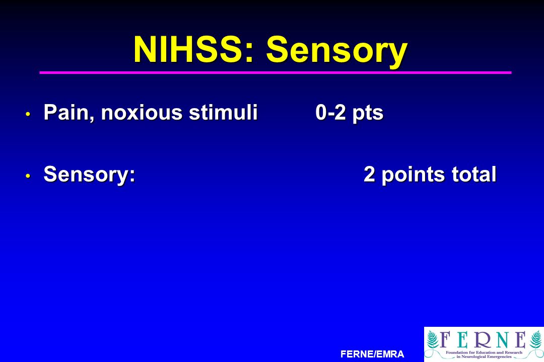
NIHSS: Sensory Pain, noxious stimuli 0-2 pts Sensory: 2 points total
54 2 54
26
NIHSS: Language Aphasia 0-3 pts Dysarthria 0-2 pts
Language: points total 54 2 54

27
NIHSS: Inattention Inattention 0-2 pts Inattention: 2 points total 54

28
NIHSS Composite CN (visual): 8 Unilateral motor: 8 LOC: 7 Language: 5
Ataxia: 2 Sensory: 2 Inattention: 2 54 2 54

29
Four Main NIHSS Areas CN/Visual: Facial palsy, gaze palsy, visual field deficit Unilateral motor: Hemiparesis LOC: Depressed LOC, poor responsiveness Language: Aphasia, dysarthria, neglect 28 total points 54 2 54

30
NIHSS ED Estimate CN (visual): 8 Unilateral motor: 8 LOC: 8
Language/Neglect: 8 Mild: 2, Moderate: 4, Severe: 8 +/- Incorporates other elements 54 2 54

31
NIHSS Patient Estimate
CN/Visual: R vision loss, no fixed gaze 4 Unilateral motor: hemiparesis 8 LOC: mild decreased LOC 2 Language: speech def, neglect 4 Approx 18 points total Moderate to severe stroke range 54 2 54

32
CT Interpretation Skills
No insular ribbon or MCA sign No detailed assessment Identify asymmetry and edema Identify blood, mass lesion Identify any area of hypodensity c/w a recent stroke of many hours duration that precludes IV tPA use 54 3 54

33
xxxx

37
IV tPA Use Skills Identify indications, contraindications
Quickly get the tests and consults Communicate with the neurologist Obtain consent with family and know what statistics are relevant Document the interaction Maintain BP below 185/110 range Follow the NINDS protocol closely

38
ED tPA Documentation With tPA, there is a 30% greater chance of a good outcome at 3 months With tPA use, there is 10x greater risk of a symptomatic ICH (severe bleeding stroke) Mortality rates at 3 months are the same regardless of whether tPA is used What was the rationale, risk/benefit assessment for using or not using tPA? What was done to expedite Rx, consult neurology and radiology early on? The key results of the NINDS trials must be explained to the patient and any family members who are assisting in the decision to use tPA. This information must be documented in the medical record. The most difficult concept to explain involves the fact that mortality rates are comparable, despite a greater intra-cranial hemorrhage rates following tPA use. This data supports the notion that a patient who has sustained an acute CVA is ill and at risk for a grave outcome,with or without tPA therapy. Once a decision has been made either to use or not to use tPA, the Emergency Medicine physician must document the rationale for the decision in terms of the potential for benefit, the inherent risks, and the preferences expressed during the discussion with the patient and their families. 54 44 54
 Mortality rates at 3 months are the same regardless of whether tPA is used. What was the rationale, risk/benefit assessment for using or not using tPA What was done to expedite Rx, consult neurology and radiology early on The key results of the NINDS trials must be explained to the patient and any family members who are assisting in the decision to use tPA. This information must be documented in the medical record. The most difficult concept to explain involves the fact that mortality rates are comparable, despite a greater intra-cranial hemorrhage rates following tPA use. This data supports the notion that a patient who has sustained an acute CVA is ill and at risk for a grave outcome,with or without tPA therapy. Once a decision has been made either to use or not to use tPA, the Emergency Medicine physician must document the rationale for the decision in terms of the potential for benefit, the inherent risks, and the preferences expressed during the discussion with the patient and their families")
39
Conclusions The IV tPA skill set is identified, limited, and manageable It is possible to provide quality emergency services with IV tPA Identify good patient candidates Make it happen quickly Document the ED management

40
The Neurological Exam in ED Stroke Patients

41
Motor Exam Is there hemiparesis & how severe? Motor: Upper & lower ext
Upper: Pronator drift, pull fingers out of hand Lower: Leg lift, hip flexion push against hand 5 17

42
Sensory Exam Is there a loss of light touch?
Sensory: Light touch, pinprick graphesthesia 5 17

43
Reflex Exam Are there pathologic reflexes? Is there a gag reflex?
Normal vs. pathologic Normal: Corneals, gag, DTRs Pathologic: Babinski, Chadduck Dec LOC, loss of airway control Loss of UMN control 5 17

44
Cerebellar Exam Is finger to nose, heel to shin OK?
Can the patient sit in the cart? Extremity motor cerebellar function Truncal ataxia and ataxic gait Positive Rhomberg 5 17

45
Visual/Neglect Exam Does the patient gaze to one side?
Is there a loss of vision on one side? Does the patient neglect one side? Persistent gaze to side of ischemic CVA Homonomous hemianopsia Neglect of one side 5 17

46
Language Exam Is the patient dysarthric?
Does the patient have an aphasia? Dysarthria: Poor mouth motor function Aphasia: Disturbed language processing Expressive: can’t speak the right words Receptive: can’t process what is heard 5 17

47
Mental Status Exam Is there an alteration in mental status?
Level of consciousness (AVPU) Alert Responds to verbal Responds to painful Unresponsive Glasgow Coma Scale Score 5 17
 Alert. Responds to verbal. Responds to painful. Unresponsive. Glasgow Coma Scale Score")
48
Case History 62 yo F with sudden onset paralysis, aphasia at 6:30 pm, no trauma No history of similar symptoms in past Patient apparently was normal prior No known risk factors (DM, HTN) No Hx surgery, bleed that would preclude tPA use 5 17
 No Hx surgery, bleed that would preclude tPA use")
49
Case Physical Exam Vital signs: Hypertension noted,
pulse ox OK, POC glucose OK HEENT: Pupils midrange, reactive, no papilledema, airway OK Neck: No Bruits, no nuchal rigidity Chest: BSBE No Rales Cardiac: No afib, no gallops or murmurs 5 17

50
Case Physical Exam (Con’t)
Abd: No evidence of AAA, peritonitis Ext: No DVT or pedal edema evident Skin: No cellulitis or wounds Neuro: Please see below 5 17

51
Case Neuro Exam CN: R mouth droop, no lid weakness
Motor: R hemiparesis, flaccid Sensory: No light touch of R extremities Reflex: No DTRs RLE, upgoing great toe R Normal corneals, normal gag reflex 5 17

52
Case Neuro Exam (Con’t)
Cerebellar: Slight truncal ataxia, to R Visual/Neglect: Lost vision & neglect, R Language: Dysarthria, expressive aphasia No receptive aphasia LOC: Slightly somnolent, responds to verbal stimuli, GCS=13 Approximate NIHSS: 18 5 17

53
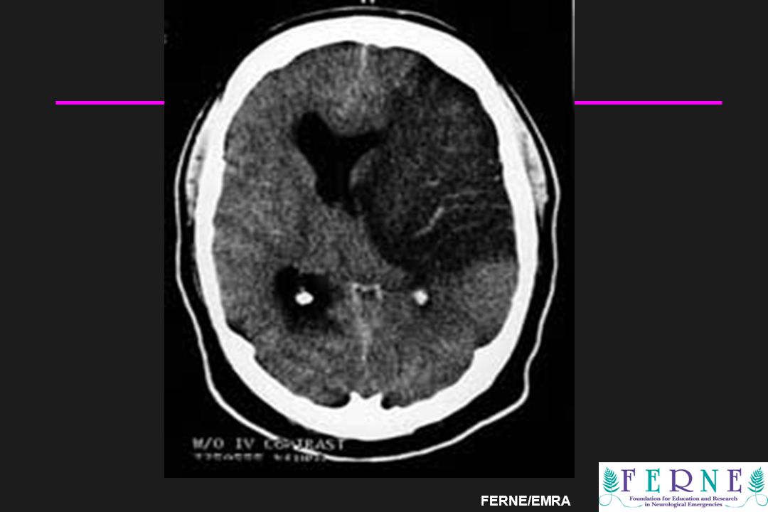
Clinical Case: CT Result
54 44 54

54
Clinical Case: ED Rx CT: no low density areas or bleed
No contraindications to tPA, BP OK NIH stroke scale: approx 18-20 Neurologist said OK to treat No family to defer tPA use tPA administered, no complications 54 44 54

55
tPA Use & Repeat Exam tPA dosing: Repeat neuro exam at 90 minutes:
8:21 pm, approx 1’45” after CVA sx onset Initial bolus: 5 mg slow IVP over 2 minutes Follow-up infusion: 40 mg infusion over 1 hour Repeat neuro exam at 90 minutes: Repeat Exam: Increased speech & use of R arm, decreased mouth droop & visual neglect Repeat NIH stroke scale: approximately 12-14 54 44 54

56
ED tPA Documentation With tPA, there is a 30% greater chance of a good outcome at 3 months With tPA use, there is 10x greater risk of a symptomatic ICH (severe bleeding stroke) Mortality rates at 3 months are the same regardless of whether tPA is used What was the rationale, risk/benefit assessment for using or not using tPA? What was done to expedite Rx and to consult neurology and radiology early on? The key results of the NINDS trials must be explained to the patient and any family members who are assisting in the decision to use tPA. This information must be documented in the medical record. The most difficult concept to explain involves the fact that mortality rates are comparable, despite a greater intra-cranial hemorrhage rates following tPA use. This data supports the notion that a patient who has sustained an acute CVA is ill and at risk for a grave outcome,with or without tPA therapy. Once a decision has been made either to use or not to use tPA, the Emergency Medicine physician must document the rationale for the decision in terms of the potential for benefit, the inherent risks, and the preferences expressed during the discussion with the patient and their families. 54 44 54
 Mortality rates at 3 months are the same regardless of whether tPA is used. What was the rationale, risk/benefit assessment for using or not using tPA What was done to expedite Rx and to consult neurology and radiology early on The key results of the NINDS trials must be explained to the patient and any family members who are assisting in the decision to use tPA. This information must be documented in the medical record. The most difficult concept to explain involves the fact that mortality rates are comparable, despite a greater intra-cranial hemorrhage rates following tPA use. This data supports the notion that a patient who has sustained an acute CVA is ill and at risk for a grave outcome,with or without tPA therapy. Once a decision has been made either to use or not to use tPA, the Emergency Medicine physician must document the rationale for the decision in terms of the potential for benefit, the inherent risks, and the preferences expressed during the discussion with the patient and their families")
57
ED tPA Documentation Patient was explained risks and benefits of tPA use and was able to understand and provide verbal consent (as able), and signature with L hand. Risk/benefit favored tPA given clear onset time, young patient with no significant morbidities or factors that would preclude tPA use, and approx NIHSS that suggests OK use. Rapid CT obtained, neurology aware of pt status, agreed with expedited tPA use, to follow. The key results of the NINDS trials must be explained to the patient and any family members who are assisting in the decision to use tPA. This information must be documented in the medical record. The most difficult concept to explain involves the fact that mortality rates are comparable, despite a greater intra-cranial hemorrhage rates following tPA use. This data supports the notion that a patient who has sustained an acute CVA is ill and at risk for a grave outcome,with or without tPA therapy. Once a decision has been made either to use or not to use tPA, the Emergency Medicine physician must document the rationale for the decision in terms of the potential for benefit, the inherent risks, and the preferences expressed during the discussion with the patient and their families. 54 44 54
, and signature with L hand. Risk/benefit favored tPA given clear onset time, young patient with no significant morbidities or factors that would preclude tPA use, and approx NIHSS that suggests OK use. Rapid CT obtained, neurology aware of pt status, agreed with expedited tPA use, to follow. The key results of the NINDS trials must be explained to the patient and any family members who are assisting in the decision to use tPA. This information must be documented in the medical record. The most difficult concept to explain involves the fact that mortality rates are comparable, despite a greater intra-cranial hemorrhage rates following tPA use. This data supports the notion that a patient who has sustained an acute CVA is ill and at risk for a grave outcome,with or without tPA therapy. Once a decision has been made either to use or not to use tPA, the Emergency Medicine physician must document the rationale for the decision in terms of the potential for benefit, the inherent risks, and the preferences expressed during the discussion with the patient and their families")
58
Hospital Course & Disposition
Hospital Course: No hemorrhage, improved neurologic function Disposition: Rehabilitation hospital 3 Month Exam: Near complete use of RUE, speech & vision improved, slight residual gait deficit Able to live at home with assistance

59
Conclusions The neurological exam can be performed in a way that is easy, understandable to other clinicians A well conducted neurological exam allows for good decisions making, and imparts a sense that the work of the emergency physician is compelling This will enhance collaboration and satisfaction, enhancing EM practice

60
Optimizing ED Ischemic Stroke Patient Care: Horizons in 2007

61
Ischemic Stroke Pt Care
Need to utilize tPA when applicable No more complicated therapeutic Risk of significant hemorrhage 50% that of imparting benefit New technologies exist Can these new diagnostics improve our ability to utilize this and other therapies? 5 17

62
Ischemic Stroke Pathophysiology
Cerebrovascular occlusion Core infarct: not salvageable Ischemic penumbra: salvageable Non-contrast CT cannot distinguish MRA and CTA may be able to 5 17

63
Diagnostics in ED CVA Pts
Core dead infarct Surrounding ischemic penumbra Non-contrast CT cannot distinguish these MRA and CTA may be able to distinguish Therapies based on whether or not there is something to salvage This enhances tPA risk/benefit profile 5 17

64
Key Clinical Questions
What do MRI and CTA/perfusion offer us when determining optimal ischemic stroke patient therapies? Which test will become our standard of care in the future? Why? 5 17

65
CNS CT, MRI : The Tests CT with contrast CT angiography (CTA)
MRI, without or with contrast MR angiography (MRA) Cerebral angiography 54 3 54
 Cerebral angiography")
66
MRI/MRA

67
Indications for MRI and CT in Emergent CNS Illness & Injury: Beyond the Non-contrast CT

68
Large, Severe CVAs Patients with acute stroke Moderate severity
NIHSS ranges from 10-20? Acute hemorrhage must be excluded Thrombolytic therapy a consideration Can pt selection be optimized? 54 3 54

69
Non-Contrast Cranial CT
Primary use is to rule out acute hemorrhage Contraindication to the use of thrombolytic therapy Identification of potential surgical candidates Limited sensitivity for detecting acute cerebral ischemia (31-75%) tPA therapy
 tPA therapy.")
70
Acute Ischemic Stroke CT
Dense MCA sign Decreased gray-white differentiation Especially in the basal ganglia Loss of insular ribbon Effacement of sulci Edema and mass effect * Large area of hypodensity* (>1/3 MCA) *May signify increased risk of hemorrhage with thrombolytic therapy
 *May signify increased risk of hemorrhage with thrombolytic therapy.")
71
Magnetic Resonance Imaging (MRI)
Multimodal MRI Demonstrates hyperacute ischemia Considered less reliable in identifying early parenchymal hemorrhage What role does MRI play in diagnosis and management of the acute stroke pt?

72
MRI: Stroke Center Approaches
CT acutely with follow-up MRI Late delineation of stroke findings Both CT and MRI acutely More expensive, time-consuming Possible enhancements in therapy? MRI acutely Is it a reasonable alternative?

73
What is Multimodal MRI? T1, T2 Imaging: Conventional weighted pulse sequences DWI: Diffusion-Weighted Imaging PWI: Perfusion-Weighted Imaging GRE: Gradient Recalled Echo pulse sequence (T2*-sensitive) FLAIR: Fluid-Attenuated Inversion Recovery images
 FLAIR: Fluid-Attenuated Inversion Recovery images.")
74
T1 & T2 Weighted Pulse Sequences
Sensitive for subacute and chronic blood Less sensitive for hyperacute parenchymal hemorrhage? Probably adequately sensitive for acute bleed

75
Gradient Recalled Echo (GRE) Pulse Sequence
May be sensitive for hyperacute parenchymal blood Detects paramagnetic effects of deoxyhemoglobin & methemoglobin as well as diamagnetic effects of oxyhgb

76
Gradient Recalled Echo (GRE) Pulse Sequence
Core of heterogeneous signal intensity reflecting recently extravasated blood with significant amounts of oxyhgb Hypodense rim reflecting blood that is fully deoxygenated

77
Diffusion-Weighted Imaging
Ischemia decreases the diffusion of water into neurons Extracellular water accumulates On DWI, a hyperintense signal Present within minutes Irreversible damage delineated Non-salvageable tissue? Infarct core

78
Perfusion-Weighted Imaging
Tracks a gadolinium bolus into brain parenchyma PWI detects areas of hypoperfusion Infarct core (DWI area) and Ischemic penumbra
 and. Ischemic penumbra.")
79
DWI/PWI Mismatch Subtract DWI signal (infarct core) from the PWI signal (infarct core and ischemic penumbra) DWI/PWI mismatch is the hypoperfused area that may still be viable (ischemic penumbra)
")
80
DWI/PWI Mismatch Important clinical implications
May identify the ischemic penumbra If there is a large mismatch, then reperfusion may be of benefit, even beyond the three hour tPA window If there is no mismatch, there may be little benefit to thrombolytic therapy, even within the three hour window

81
DWI/PWI Mismatch PWI hypoperfused area DWI signal

82
So what is the role of MRI in the ED evaluation of the stroke patient?
Secondary? Initial CT to rule out hemorrhage Subsequent MRI to fully delineate ischemia, infarct and to follow treatment Primary? Initial and possibly only imaging modality

83
MRI in Large, Severe CVAs
Primary MRI not current EM standard Logistical, timing issues exist MRI likely able to diagnose hemorrhage DWI/PWI mismatch a promising exam Tailored thrombolytic therapy?? Improved patient outcome?? 54 3 54

84
CT Angiography & CT Perfusion

85
CT Angiography and CT Perfusion
Essential questions Is there hemorrhage? Is there large vessel occlusion? Is there “irreversibly” infarcted core? Is there “at risk” penumbra? One contrast bolus yields two datasets Vessel patency Infarct versus salvageable penumbra

86
CT Angio & Perfusion

87
CT Perfusion Terminology
Blood Flow Blood Volume Mean Transit Time or Time to Peak

88
Case: Value of CTA/CTP within 3 hour window
50 yo male CT within hour of symptom onset Awake, alert, dysarthric Fixed right sided gaze Left sided weakness Initial

89
Case: Value of CTA/CTP within 3 hour window

90
Case: “Wake up” Stroke 0735 at outside hospital

91
Case: “Wake up” Stroke

92
Case: “Wake up” Stroke 1030 at stroke center

93
Case: “Wake up” Stroke 24 hours later at stroke center

94
Conclusions Diagnostics may guide future therapies, esp when onset time and penumbra size uncertain May be able to maximize benefit and minimize risk through greater understanding of infarct core and salvageable ischemic penumbra Future CTA use like non-contrast CT use today Software for rapid reconstruction exists MRI/MRA still has too many technical hurdles EM physicians need to consider next steps 5 17

95
Treating Intracerebral Hemorrhage in the Anti-coagulated Patient

96
Clinical History 66 year old male presents with acute onset of aphasia and right sided weakness while eating at home Initially complained of a headache BP of 220/118 mm Hg Accucheck 316 Initial GCS of 14 54 3 54

97
Paramedic’s Report Patient less responsive than initially
Aphasia and weakness worsening? He is on a “bag o’ meds” Per family, started an antibiotic a week ago 54 3 54

98
ED Presentation ED VS BP 224/124, P 100, RR 16, T 98.8, pulse ox 99% Somnolent, but slowly responds to simple commands Snores a bit when not stimulated Clear lungs and a regular cardiac rate and rhythm Neurological screening exam Pupils midpoint, equal and reactive L sided gaze preference R facial weakness R upper > lower extremity weakness Expressive aphasia 54 3 54

99
Key Clinical Questions
What are the key diagnostic issues? What are the potential complicating factors? What guidelines direct potential therapies? What is the urgency of potential interventions? What is the relative availability of those therapies in our institution? 54 2 54

100
Bag o’ Meds

101
The Great American Poison

102
Which of these belong to this patient?

103
Elevated INR Therapy: The Procedure

104
INR Based on the Prothrombin time test
Sensitive to reductions of Vitamin-K dependent clotting factors II, VII, and X Not factor IX Designed specifically for stably anticoagulated patients May be inappropriate test following replacement therapy with either plasma or clotting factor concentrates

105
Elevated INR Rx Procedure
Vitamin K 10 mg by slow IV infusion 54 2 54

106
Vitamin K Necessary to achieve more than a temporary reversal of anticoagulation Adequate response requires at least 2-6 and up to 24 hours Anaphylactic or anaphylactoid reactions rarely associated with IV administration Safest and most rapidly acting route of administration unclear Wjasow C, McNamara R. J Emerg Med 2003;24(2): Fiore LD et al. J Thrombosis & Thrombolysis 2001;11(2):
: Fiore LD et al. J Thrombosis & Thrombolysis 2001;11(2):")
107
Coagulation Factor Replacement
Options include FFP Prothrombin Complex Concentrates (PCC) Recombinant Factor VIIa Normal coagulation achieved more rapidly with PCC, rFVIIa than with FFP Fredriksson K et al. Stroke 1992;23: Makris M et al. Thromb Haemostasis 1997;77:
 Recombinant Factor VIIa. Normal coagulation achieved more rapidly with PCC, rFVIIa than with FFP. Fredriksson K et al. Stroke 1992;23: Makris M et al. Thromb Haemostasis 1997;77:")
108
Bedside Realities: Can you answer these process questions?
Is thawed FFP immediately available from your blood bank? How long will it take your blood bank to get it to you? Does your hospital blood bank or inpatient pharmacy store PCC and rFVIIa? What is the relative rapidity of response of each of these agents?

109
Elevated INR Rx Procedure
Vitamin K 10 mg by slow IV infusion Fresh frozen plasma (5-8 ml/kg, 1-2 units, cc total) 54 2 54
")
110
Elevated INR Rx Procedure
Vitamin K 10 mg by slow IV infusion Fresh frozen plasma (5-8 ml/kg, 1-2 units, cc total) Prothrombin Complex Concentrate IU/kg Dose based on Factor IX units Alternatively, 500 IU initially followed by second administration of 500 IU according to the INR value measured just after the first administration OR 54 54 2
 Prothrombin Complex Concentrate IU/kg. Dose based on Factor IX units. Alternatively, 500 IU initially followed by second administration of 500 IU according to the INR value measured just after the first administration. OR")
111
Elevated INR Rx Procedure
Vitamin K 10 mg subq or IVP Fresh frozen plasma (5-8 ml/kg) 1-2 units, cc total Prothrombin Complex Concentrate IU/kg Recombinant Factor VIIa (40-60 µgr/kg) Usually 3-4 mg total OR OR 54 54 2
 1-2 units, cc total. Prothrombin Complex Concentrate IU/kg. Recombinant Factor VIIa (40-60 µgr/kg) Usually 3-4 mg total. OR. OR")
112
Drawbacks to FFP Reversing OAC
Time-consuming? Can delay neurosurgical evacuation May require clinically substantial IV fluid volumes Contains a variable content of Vitamin K-dependent clotting factors May not completely correct INR May not adequately correct for factor IX Risk of viral transmission Not pooled HIV ≈ 1:1,900,000 Hepatitis C ≈ 1:1,000,000 Hepatitis B ≈ 1:137,000 Makris M et al. Thromb Haemostasis 1997;77:

113
PCC Prepared from pooled plasma of thousands of blood donors
Less viral transmission risk than FFP Contains vitamin K-dependent procoagulant and factors Infused over 15 minutes Relative thromboembolic risk unclear Acquisition cost of usual adult dose ≈ $450 Abe et al. Rinsho to Kenkyu [in Japanese] 1987;64: Sorensen B et al. Blood Coagulation and Fibrinolysis 2003:14:

114
Recombinant Factor VIIa
Rapid onset of action Almost immediate Clinically apparent hemostasis in 10 minutes Short half life (2.3 hours) Relatively high acquisition cost ≈ $2,500-$3,500 for 3-4 gm dose Park p et al. Neurosurgery 2003;53:34-39. Sorensen B et al. Blood Coagulation and Fibrinolysis 2003:14: Novoseven [package insert]. Princeton, NJ: Novo Nordisk Pharmaceuticals, Inc; 2003.
 Relatively high acquisition cost. ≈ $2,500-$3,500 for 3-4 gm dose. Park p et al. Neurosurgery 2003;53: Sorensen B et al. Blood Coagulation and Fibrinolysis 2003:14: Novoseven [package insert]. Princeton, NJ: Novo Nordisk Pharmaceuticals, Inc;")
116
ED Treatment and Patient Outcome

117
ED Patient Management The BP treated with IV labetalol
The INR was noted to be 5.6 Vitamin K administered 2 units FFP administered Pt admitted to the neurosurgical ICU 54 3 54

118
Patient Outcome The hemorrhage size increased slightly on CT with slight intraventricular extension The patient’s clinical condition slightly improved gradually Discharged to rehab 10 days after admission 54 3 54

119
ED ICH Patient Rx: A Retrospective

120
OAC Related ICH Know the treatment guidelines
Know the relative availability at your institution of different coagulation factor replacements Communicate with neurosurgical consultants regarding a potential indication for PCC or rFVIIa use

121
Conclusions Ischemic stroke and ICH are EM diseases
Treatment options are easy to identify Standards can be met Pt outcomes can be optimized EM clinical practice can be optimized

122
Thank you. www.ferne.org ferne@ferne.org edsloan@uic.edu 312 413 7490
ferne_ema_2007_neuroresus_strokeich_sloan_092407_finalcd 4/5/ :53 PM Edward P. Sloan, MD, MPH, FACEP 54 54 1

123
54 1 54
Similar presentations


 in Emergency Department Patients with Acute Stroke Edward Sloan, MD, MPH Professor Department of Emergency Medicine.>")


