Download presentation
Presentation is loading. Please wait.

1
Brain Tumours Mostafa EL-Haddad
M.B.B.ch., Msc., MD., FRCR(UK)., Kasr El-Ainy Hospital Cairo University (NEMROCK) 2012
., Kasr El-Ainy Hospital Cairo University (NEMROCK)")
2
Genetic Syndromes

3
Syndrome Tumors Other Asso. Genetics -Turcot’s - Glioblast. - Medullobla. - Colon ca APC gene (5q) A. Dominant -Tuberous Sclerosis -Subependymal giant cell astrocytoma. -Hamartomas. -Angiofibromas. -Hypomelanotic patches. -M retardation. -TSC1 and TSC2 -9q and 16p. -A.D NF I (commonest of these rare syndromes) -Neurofibromas. -Gliomas. -Sarcomas. -Café au lait patches. Lish nodules. Neural crest tumors. NF I Ch 17. A.D. NF II Meningiomas. Shwannomas (AN) Gliomas(spinal mainly) Lens opacity. Cerebral calcifications NF2 on ch22
 -Neurofibromas. -Gliomas. -Sarcomas. -Café au lait patches. Lish nodules. Neural crest tumors. NF I Ch 17. A.D. NF II. Meningiomas. Shwannomas. (AN) Gliomas(spinal mainly) Lens opacity. Cerebral calcifications. NF2 on ch22.")
4
VHL g Syndrome Tumors Other Asso. Genetics Cowden
Dysplastic gangliocytoma of cerebellum -Peripheral hamartomas. -Breast ca. -Thyroid ca -Colorectal ca. PTEN/MMAC1 10q. A.D. Basal Naevus (Gorlin’s) -Medulloblas. Basal cell cas. Bone ablnormalities Palmar pits PTCH ch 9q. Von-Hipple lindau Cerebral and spinal haemangioblastomas Wilm’s VHL g Li-Fraumeni -Gliomas. -PNETS -Sarcomas. Breast. P53.
 -Medulloblas. Basal cell cas. Bone ablnormalities. Palmar pits. PTCH ch 9q. Von-Hipple lindau. Cerebral and spinal haemangioblastomas. Wilm’s. VHL g. Li-Fraumeni. -Gliomas. -PNETS. -Sarcomas. Breast. P53.")
5
Infratentorial tumors more common in children (infants)
Infratentorial tumors more common in children (infants). Also in midline. in adults Supratentorail more.
. Also in midline. in adults Supratentorail more.")
6
Sex In general male more. Meningioma and Shwannomas more in female
Ependymoma and nerve sheath equally distributed.

7
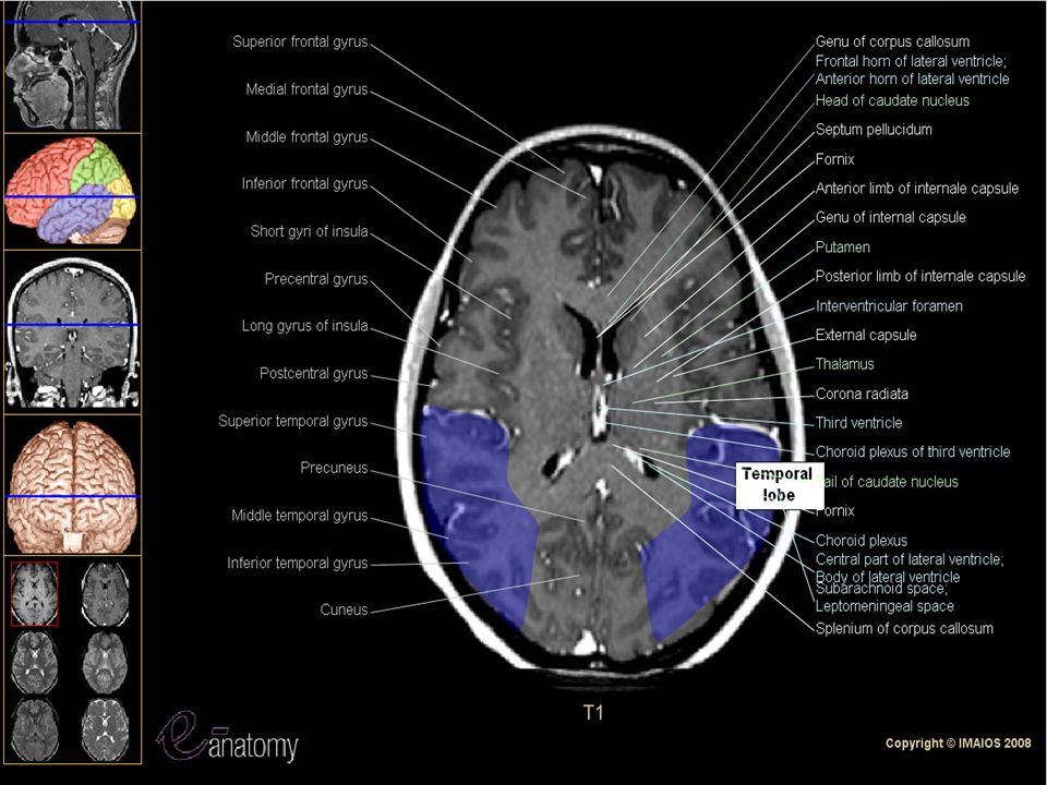
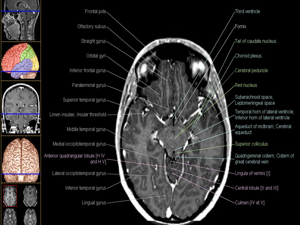
Anatomy Circle of Willis. Optic Chiasma.
The frontal and temporal lobes are separated by the Sylvian fissure, while the parietal and occipital lobes are separated by the calcarine sulcus

8
ANATOMY

29
Staging AJCC 2002: NO Old TNM M staging and medulloblastoma
Biopsy no biopsy.

30
Commonest Brain Tumor? Brain Metastasis.

31
Imaging T2 and FLAIR (fluid-attenuated inversion recovery) images are more sensitive for detecting edema. For nonenhancing tumors, especially glial neoplasms, the FLAIR sequence and T2 sequences are best for the definition of tumor extent. CSF spread of tumor may be associated with several abnormal findings by CSF examination. These include CSF pressure above 150 mm H2O at the lumbar level in a laterally positioned patient, elevated protein level, typically >40 mg/dL, a reduced glucose level (below 50 mg/mL), and the finding of tumor cells by cytologic examination. Tumor markers in the CSF may help in making the diagnosis.
, and the finding of tumor cells by cytologic examination. Tumor markers in the CSF may help in making the diagnosis.")
32
WHO Grading Classification idea: -Neuroepithelial. -Germ cells.
-Meningeal . -Etc……… OLIGODENDROGLIOMA WHY SPECIAL?

33
Brain Alpha-Beta ratio
Implication in Dose and fractionation.

34
General Management Newer generation anticonvulsants, such as levetiracetam, lamotrigine, and pregabalin that do not affect cytochrome P450 activity are now preferred.

35
Choose Your Patient Low Grade. High grade. Children.
Boost or No Boost? Dose escalation in low and High grade gliomas. Risk structures.

36
CT-Simulator Patient preparation. IV contrast.
Treatment position: Supine? Prone?. Neck flexed or extended .WHEN? Fixation methods Supine:less movement with respiration, more comfortable. Prone: push the intestine up and the seminall vesicle forward away from the rectum.

37
Define your target Margin for Low grade tumors
Margin in High grade tumors.

38
GTV A central low-density area is evaluated as necrosis and is surrounded by an annular enhancing area corresponding to a densely cellular zone of viable neoplasm called the “rim.” (IF any) The external limit of the rim corresponds to the GTV
 The external limit of the rim corresponds to the GTV.")
39
GTV In High Grade Glioma
MRI or CT? both with contrast. Best with T1 MRI with contrast.

40
GTV in Low grade Glioma CT in Low grade glioma, tumor is non enhancing so its very difficult to delineate. MRI T1 or T2?

41
CTV in High Grade Glioma
2-3 cm beyond any existing edema. Which imaging study will you use to adequately define your edema?? MRI ?? T1 or T2? .

42
Brain Metastasis

45
Brain Metastasis RPA Class I KS≥70. Primary: controlled. Age ≤65.
No extra-cranial metastasis. MS 7.1 RPA Class II KPS ≥70 Primary: Uncontrolled And/or Age ≥ 65 And/or Extra-cranial metastasis MS 4.3 RPA Class III KPS <70 MS 2.3 Gaspar L, Scott C, Rotman M, et al. Recursive partitioning analysis (RPA) of prognostic factors in three Radiation Therapy Oncology Group (RTOG) brain metastases trials. Int J Radiat Oncol Biol Phys. 1997;37: Gaspar et al IJROBP 1997
 of prognostic factors in three Radiation Therapy Oncology Group (RTOG) brain metastases trials. Int J Radiat Oncol Biol Phys. 1997;37: Gaspar et al IJROBP")
46
Factors NOT in The RTOG Prognostic Factors
Number of the lesions. Primary site (breast, lung…). Time interval between the Metastasis and the primary. Location. Neurologic Function. Radiation dose. Tumor response.
. Time interval between the Metastasis and the primary. Location. Neurologic Function. Radiation dose. Tumor response.")
47
Karnofsky scale 100%: Normal no complaints; no evidence of disease
90%: Able to carry on normal activity; minor signs or symptoms of disease. 80%: Normal activity with effort; some signs or symptoms of disease. 70%: Cares for self; unable to carry on normal activity or to do active work. 60%: Requires occasional assistance, but is able to care for most of his personal needs.

48
50%:Requires considerable assistance and frequent medical care.
40%: Disabled; requires special care and assistance. 30%:Severely disabled; hospital admission is indicated although death not imminent. 20%: Very sick; hospital admission necessary; active supportive treatment necessary. 10%: Moribund; fatal processes progressing rapidly. 0 Dead

49
Value of Whole Brain Irradiation
Increase Median Survival from 1 to 4 months.

50
Radio-surgery When? RTOG 9508. RPA class III. RPA class I.
Single Mets. Andrews DW, Scott CB, Sperduto PW, et al. Whole brain radiation therapy with or without stereotactic radiosurgery boost for patients with one to three brain metastases: Phase III results of the RTOG 9508 randomised trial. Lancet. 2004;363: Patients in RPA class 3 were excluded. Patients with single brain metastasis treated with RS survived longer (6.5 months vs. 4.9 months, p 0.04) than those treated with WBRT alone. However, there was no survival benefit from adding RS for patients with multiple metastases. Additional analysis found a notable survival advantage from RS in patients in RPA class 1 regardless of the number of brain metastases (11.6 vs. 9.6 months), and in patients whose largest tumor was more than 2 cm in diameter or who exhibited either squamous histology or non-small cell lung cancer. Pa- tients treated with RS had markedly better performance status and decreased corticosteroid use at 6 months com- pared to the control arm. Moreover, as expected, RS did improve local response rates and time to local failure. These results pertained even though 19% of patients in the RS arm never received RS. Nonetheless, there was no difference in time to intracranial failure or neurological death rates. The authors concluded that RS boost to WBRT should become standard for patients with a single metastasis,; and because of its favorable impact on perfor- mance status, it also should be considered for those patients with two or three metastases.11 treating patients’ brain metastases principally with radiosurgery, deferring whole brain radiother- apy because of concerns regarding neurotoxicity
 than those treated with WBRT alone. However, there was no survival benefit from adding RS for patients with multiple metastases. Additional analysis found a notable survival advantage from RS in patients in RPA class 1 regardless of the number of brain metastases (11.6 vs. 9.6 months), and in patients whose largest tumor was more than 2 cm in diameter or who exhibited either squamous histology or non-small cell lung cancer. Pa- tients treated with RS had markedly better performance status and decreased corticosteroid use at 6 months com- pared to the control arm. Moreover, as expected, RS did improve local response rates and time to local failure. These results pertained even though 19% of patients in the RS arm never received RS. Nonetheless, there was no difference in time to intracranial failure or neurological death rates. The authors concluded that RS boost to WBRT should become standard for patients with a single metastasis,; and because of its favorable impact on perfor- mance status, it also should be considered for those patients with two or three metastases.11 treating patients’ brain metastases principally with radiosurgery, deferring whole brain radiother- apy because of concerns regarding neurotoxicity.")
51
Can we avoid WBI? EORTC: 22952 –26001.
The American Surgeons Oncology Group. Japanese Radiation Oncology Study Group. The third such study, from the Japanese Radiation Oncology Study Group (JROSG 99-1), randomly assigned patients with one to four brain metastases to RS with or without WBRT (30 Gy in 10 fractions). Overall survival was the primary endpoint, with cause of death, intracra- nial recurrence, and toxicity as secondary endpoints. The study closed prematurely when interim analysis indicated that more than 800 patients would be required to demon- strate a significant difference in the primary endpoint. The median survival was identical in the two arms (7.5 months for RS alone and 8.0 months with WBRT). Unsur- prisingly, intracranial failure was lower at 1 year in the WBRT arm (46.8%) compared with the RS-alone arm (76.4%). There was no difference in the percentage of deaths attributed to neurologic progression between the two groups. The authors concluded that the addition of WBRT did not improve survival but decreased intracra- nial relapse and the need for salvage treatment.20 The study has been criticized for being vastly underpowered for a survival end point.WBRT became the standard of care,25 with a rate of local relapse of 10% to 20%.
, randomly assigned patients with one to four brain metastases to RS with or without WBRT (30 Gy in 10 fractions). Overall survival was the primary endpoint, with cause of death, intracra- nial recurrence, and toxicity as secondary endpoints. The study closed prematurely when interim analysis indicated that more than 800 patients would be required to demon- strate a significant difference in the primary endpoint. The median survival was identical in the two arms (7.5 months for RS alone and 8.0 months with WBRT). Unsur- prisingly, intracranial failure was lower at 1 year in the WBRT arm (46.8%) compared with the RS-alone arm (76.4%). There was no difference in the percentage of deaths attributed to neurologic progression between the two groups. The authors concluded that the addition of WBRT did not improve survival but decreased intracra- nial relapse and the need for salvage treatment.20 The study has been criticized for being vastly underpowered for a survival end point.WBRT became the standard of care,25 with a rate of local relapse of 10% to 20%.")
52
Landmarks Sella turcica is centrally located and marks the lower border of the median telencephalon and diencephalon. Reid's baseline and the Frankfort horizontal plane. Optic canal runs at most 1 cm superior and 1 cm anterior to that point. Pineal body 1 cm posterior and 3 cm superior to the external auditory meatus. plane:connecting points in the two external auditory meatus and one anterior infraorbital edge)
")
53
What’s FLAIR? Think of the FLAIR sequence as a bit of a 'negative', in that FLAIR will show areas of differing fluid concentration (not blood). FlAIR are modified T2-weighted sequences on which fluid in motion, such as cerebrospinal fluid, remains dark, whereas tumor or edema remains white. This sequence is useful for the delineation of periventricular lesions. The areas of high fluid concentration are an indication of edema (swelling), and might (emphasis on might), indicate an area of concern. Typically when something starts growing in the olde brain bucket, it causes swelling. FLAIR provide information about water motion and can be helpful for differentiation between vasogenic (especially in strokes) and cytotoxic edema or for the differential diagnosis between tumor or abscess
, and might (emphasis on might), indicate an area of concern. Typically when something starts growing in the olde brain bucket, it causes swelling. FLAIR provide information about water motion and can be helpful for differentiation between vasogenic (especially in strokes) and cytotoxic edema or for the differential diagnosis between tumor or abscess.")
54
MR Spectroscopy MR spectroscopy is a promising approach for both metabolic and functional evaluation of GTV and CTV. This technique provides information about tumor activity based on the levels of cellular metabolites.

55
Choline is a neurotransmitter and membrane component that is increased in glioma and the degree of elevation of the choline level correlates with elevation of tumor cell density. N-acetylaspartate is a neuronal metabolite that is decreased in glioma implying an increase in the choline-to-N-acetylaspartate ratio.

56
Creatine indicates a cellular energetic process, and lactate is a catabolite of anaerobic metabolism that can be used as a surrogate marker for necrosis or hypoxia.

57
CT and MRI delineation of GTV and CTV for untreated glioma remains a controversial and difficult issue, mainly because of the discrepancy between real tumor invasion and that estimated by CT or MRI.

60
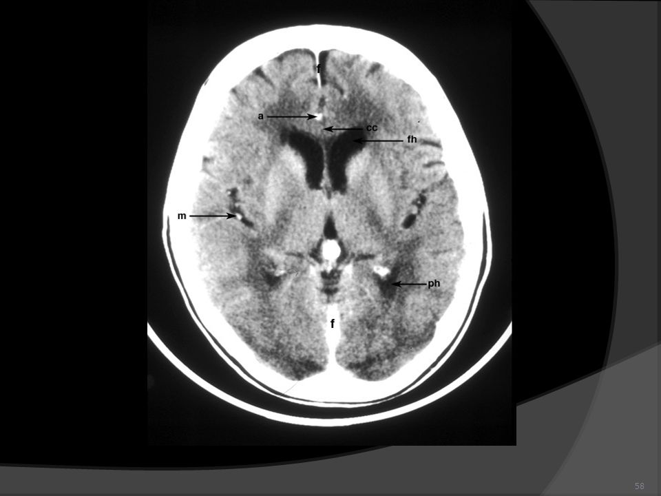
Cranial structures Hard Palate Nasopharynx Sphenoid air sinus
Pituitary gland Frontal sinus Frontal lobe Corpus callosum Septum pellucidum Parietal lobe Fourth ventricle Occipital lobe Cerebellum Sinus Confluence Pons Medulla Oblongata Spinal Cord

61
High Grade Tumors Grade 3: Anaplastic astrocytoma and pure and mixed anaplastic oligodendroglioma Grade 4: Glioblastoma Multiforme

62
Survival in Months Anaplastic Oligodendroglioma ~60.
Anaplastic Astrocytoma ~36. Glioblastoma Multiforme < 12.

63
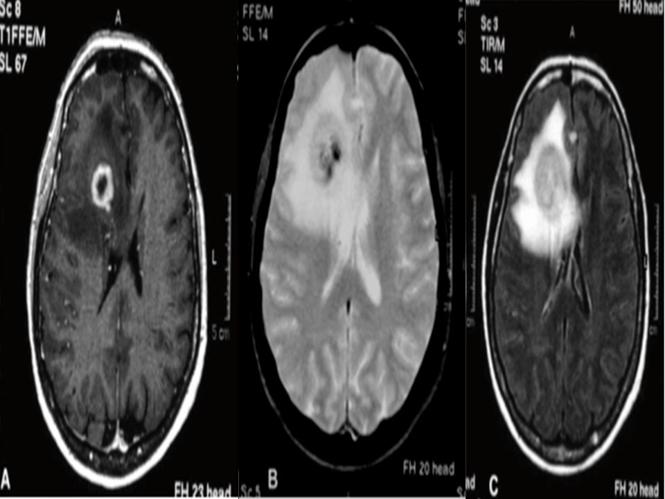
Grade 4 Astrocytoma 6 cm right posterior parietal lesion
Irregular margins, infiltrating tumour Rim-enhancing, central necrosis Mass effect / edema Beware corpus callosum involvement

64
High grade Astrocytoma: Differential Diagnosis
Vs abscess

65
High grade astrocytoma: DD
Abscess. Metastasis. Resolving hemorrhagic stroke. multiple sclerosis, giant plaque. Early-delayed radiation edema can mimic tumour progression

66
Glioblastoma Multiforme:
80% of all malignant glioma. Median survival is 9-10 months. 5 yr survival < 5%. Rarely metastasize. >80% failures within 2 cm of original tumor. Calcification occurs only when the glioblastoma arose from a low-grade.

67
Types of GBM 1ry: 2nd:

68
Prognostic Factors (for HG Gliomas)
RTOG (Curran.1993) Age Histology KPS Mental status Duration of symptoms Neurologic functioning class, Extent of surgery Radiation dose All significant predictive factors of outcome. favorable group (classes I – II): 12% of all patients; 2YOS of ~ 70% intermediate group (classes III – IV): 43% of all patients; 2YOS of 10 – 30% unfavorable group (classes V – VI): 45% of all patients; 2YOS of ~ 5%
 Age. Histology. KPS. Mental status. Duration of symptoms. Neurologic functioning class, Extent of surgery. Radiation dose. All significant predictive factors of outcome. favorable group (classes I – II): 12% of all patients; 2YOS of ~ 70% intermediate group (classes III – IV): 43% of all patients; 2YOS of 10 – 30% unfavorable group (classes V – VI): 45% of all patients; 2YOS of ~ 5%")
70
RPA For GBM Class III Age: Tumor Type: Mental Status:
Performance status: < 50 GBM KPS

71
RPA For GBM Class IV Age: Tumor Type: Mental Status:
Performance status: < 50 GBM KPS <90

72
RPA For GBM Class IV ≥50 GBM Good neurologic function
Age: Tumor Type: Mental Status: Performance status: Treatment status : ≥50 GBM Good neurologic function Surgical resection

73
RPA For GBM Class V Treatment status : Age: Tumor Type: Mental Status:
≥50 GBM Neurologic function that inhibits the ability to work 70-100 Surgical resection or biopsy only followed by at least 54.4 Gy radiotherapy Good neurologic function Surgical resection Age: Tumor Type: Mental Status: Performance status: Treatment status :

74
RPA For GBM Class V Age: Tumor Type: Mental Status:
≥50 GBM Normal <70 Age: Tumor Type: Mental Status: Performance status:

75
Why The Tumor Enhance? What decrease the enhancement? What increase?
The contrast enhancement that high lights a glioblastoma multiforme on these scans results from dye extravasating through the disrupted blood-brain barrier (BBB) Glioblastoma Multiforme Glucocorticoids decrease by increasing BBB integrity, radiation or steroid withdrawal may increase by increasing BBB distruption,
 Glioblastoma Multiforme. Glucocorticoids decrease by increasing BBB integrity, radiation or steroid withdrawal may increase by increasing BBB distruption,")
76
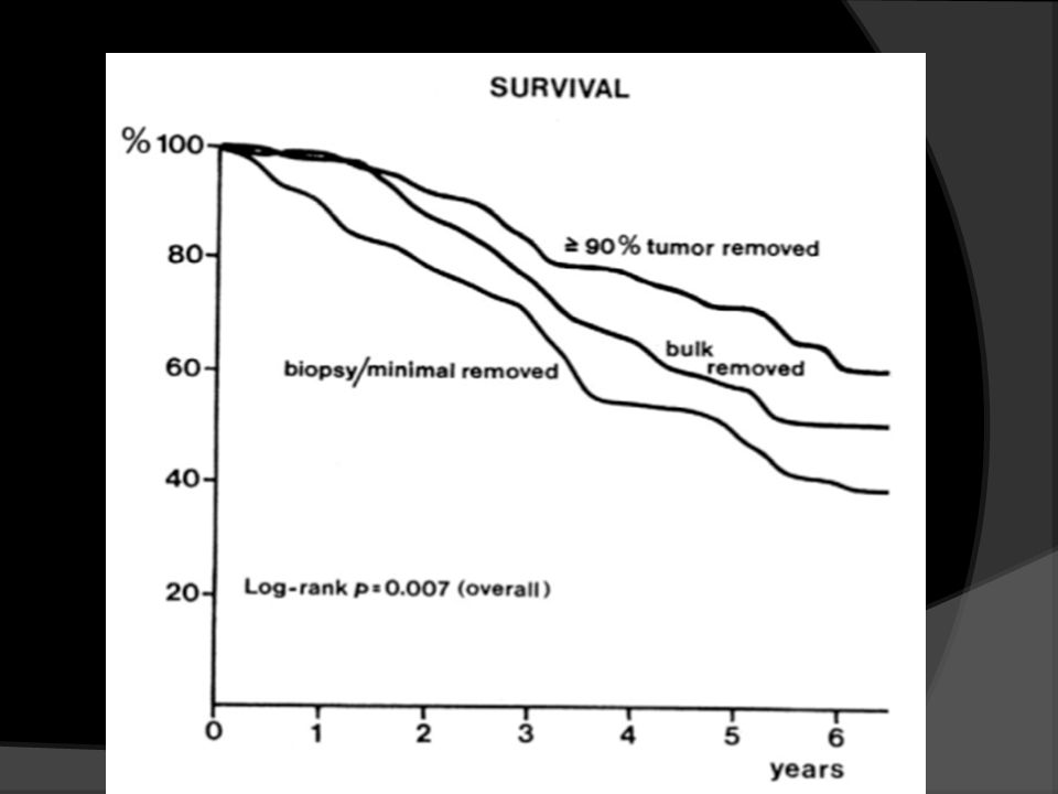
Treatment Surgery: Controversy exists regarding the relationship between the extent of surgery and survival. in this patient population, there is uniform agreement that optimal treatment of glioblastoma multiforme involves removing as much tumor as possible without causing neurologic deficits. Grossman R, et al: National Comprehensive Cancer Network adult brain tumor practice guidelines. Oncology 11: , 1997.

77
Partial Brain 60Gy is The Standard
Radiotherapy Initially 50 to 60Gy of whole-brain radiation following surgery significantly increases the median survival of patients with malignant gliomas from 14 to 36. Partial Brain 60Gy is The Standard

78
Les études postmortem montrent que dans l’œdème, le risque de présence de cellules tumorales est surtout important dans les 20 mm avoisinant le volume tumoral macroscopique. , cette distance de 20 mm correspondant à la marge de sécurité habituellement réaliséeLe risque d’extension méningée de contiguïté est plus facilement évalué pour les formes corticales par un examen par résonance magnétique

79
Le risque d’extension pour les formes profondes dans les noyaux gris et surtout les fibres longues associatives (corps calleux et capsule) est important et peu évalué. En cas de volumineuse tumeur hémisphérique à proximité du corps calleux, le dépassement de la ligne médiane se jus-Volume tumoral macroscopique (GTV) En cas de tumeur nodulaire (surtout de type II), le volume tumoral macroscopique correspond à la zone d’hypoden- sité tomodensitométrique, d’hyposignal en mode T1 ou
 En cas de tumeur nodulaire (surtout de type II), le volume tumoral macroscopique correspond à la zone d’hypoden- sité tomodensitométrique, d’hyposignal en mode T1 ou.")
80
The exact volume of treatment is controversial and may be based either on contrast- enhancing disease or T2 changes with a margin or a combination of both.

81
Unit A Protocol A typical treatment schema might include 45 to 46Gy administered to T2 changes on MRI with a 1- to 2-cm margin, with an additional boost of 14 to 15Gy to the area of contrast enhancement with a margin.

82
Edema or NOT

84
Temozolamide ( 6cycles ). ASCO 2004 NEJM (Stupp et al)
RCT (concomitant + adjuvant TMZ and RT versus RT alone) RT (6000 cGy in 30 fractions - retina limited to 50 Gy, brainstem and chiasm limited to 54 Gy) TMZ (concomitant) 75 mg/m2 (weekly CBC; Septra prophylaxis for PCP-consider for lymphocyte count <500) TMZ (adjuvant) mg/m2 daily x 5 d, q 28 d ( 6cycles ).
 RT (6000 cGy in 30 fractions - retina limited to 50 Gy, brainstem and chiasm limited to 54 Gy) TMZ (concomitant) 75 mg/m2. (weekly CBC; Septra prophylaxis for PCP-consider for lymphocyte count <500) TMZ (adjuvant) mg/m2 daily x 5 d, q 28 d. ( 6cycles ).")
85
Irradiation Volume in Stupp Trial
GTV plus 2 to 3 tumor margin for CTV.

87
MGMT Story

88
LAW BAwaZ MYNE YENKEZ DNA
Temozolamide YEBAWAZOH MGMT DNA YENKEZOH YEMOUT LAW BAwaZ MYNE YENKEZ DNA

91
Adjuvant Chemotherapy
Rules of Adjuvant Treatment: 1- All known tumor should be removed. 2- Effective chemotherapy must be available 3- Chemotherapy Start as soon as possible after surgery Give at maximum tolerated dose Continue for limited time

92
Define Parinauds syndrome
Decreased upward gaze Dissociation near-light (i.e., limited restriction to light but retained response to accommodation) Diminished convergence caused by pressure into superior colliculi and lateral geniculate nuclei. Other signs of pineal gland tumor: diplopia, visual defect, papilledema, ↓visual activity
 Diminished convergence. caused by pressure into superior colliculi and lateral geniculate nuclei. Other signs of pineal gland tumor: diplopia, visual defect, papilledema, ↓visual activity.")
93
Hypofractionation schedules are also used in selected patients
Hypofractionation schedules are also used in selected patients. This approach shortens the treatment time without substantially compromising survival. However, as hypofractionation is likely to in- crease the late effects of radiation, it is usually limited to patients with advanced age or poor performance status, whose life ex- pectancy is very limited.

96
Treatment Challenges The New Approaches to Brain Tumor Therapy (NABTT) CNS Consortium used paclitaxel in a dose and schedule that caused myelosuppression and alopecia in patients with breast cancer and lymphomas.13 However, no significant tox- icities were observed in this glioblastoma multiforme trial, and paclitaxel levels were noted to be much lower than seen in systemic cancers. It was subsequently recognized that all of these patients were taking phenytoin, a potent P450 enzyme inducer, and that increased hepatic metabolism of paclitaxel was responsible for the low levels and lack of observed tox- icities.

97
Meningioma Imaging: MRI? CT which is better?

98
WHO Classification Grade I or benign meningiomas:
low mitotic index (90% of all meningiomas). Grade II or atypical meningiomas: higher mitotic index (>4 mitosis per 10 HPF) (5%–7% of all meningiomas). Grade III or malignant meningiomas: highest mitotic index (>20 per 10 HPF) (1%–3% of all meningiomas).
. Grade II or atypical meningiomas: higher mitotic index (>4 mitosis per 10 HPF) (5%–7% of all meningiomas). Grade III or malignant meningiomas: highest mitotic index (>20 per 10 HPF) (1%–3% of all meningiomas).")
100
Grade I meningioma does not metastasize.
Rarely grade II and III meningioma may metastasize outside of the central nervous system, especially in advanced or recurrent diseases.

101
Treatment Watch and Wait: small asymptomatic.
Surgery: Complete is the Goal. Completeness of resection can be determined by early (<72 h) postoperative, contrast-enhanced CT scan or MRI of the brain.
 postoperative, contrast-enhanced CT scan or MRI of the brain.")
102
Simpson Criteria The 10-year recurrence rate is 9% for patients with a Simpson Grade of 1, compared to a 10-year recurrence rate of 29% for patients with a Simpson grade of 3 (Level IV) (Simpson 1957).
 (Simpson 1957).")
103
Simpson Criteria

104
GI incomplete resection
GI completely resected GI incomplete resection Radiotherapy GII or GIII

105
Partial resection plus RT equal to complete resection.

106
Technique The gross tumor volume (GTV) should be determined by fusion of the T1-weighted contrast- enhanced MRI with the planning CT (Grade B). The tumor volume is best determined by comparing preoperative and postoperative findings of a contrast-enhanced MRI of the brain.

107
MRI defined meningioma volumes could be larger but not inclusive of CT defined volumes, thus CT scan and MRI are complementary for the purpose of tumor delineation (Khoo et al. 2000).
..")
108
Adjuvant radiation therapy The GTV for adjuvant radiation of WHO Grade I meningioma can include the postoperative residual tumor only. For atypical and malignant meningioma (i.e., WHO grade II and III), GTV should include the entire tumor bed on the pre- operative MRI. Dose: 50 to 54Gy for GI. Up to 60 for GII and III.
, GTV should include the entire tumor bed on the pre- operative MRI. Dose: 50 to 54Gy for GI. Up to 60 for GII and III.")
109
Meningioma and Hormonal Therapy
Approximately 70% of meningiomas are progesterone receptor positive and 30% are estrogen receptor positive (Sanson 2000). Should we use it??
. Should we use it")
110
Biotherapy and Chemotherapy
FASHAL !! Somatostatin under investigatioins

113
Low grade Gliomas

114
LOW GRADE GLIOMAS Grade (based on): 1. nuclear abnormalities
2. Mitoses 3. endothelial proliferation 4. necrosis. Mayo system adds the number of features found above. I=0, II=1, III=2, IV=3-4. Kernohan is another major grading system. PATHOLOGY (WHO) Astrocytic Tumors Astrocytomas 70% fibrillary protoplasmic gemistocytic Pilocytic astrocytomas Pleomorphic xanthoastrocytomas Subependymal giant cell astrocytomas Oligodendroglial Tumors Mixedoligoastrocytoma
 Astrocytic Tumors. Astrocytomas 70% fibrillary. protoplasmic. gemistocytic. Pilocytic astrocytomas. Pleomorphic xanthoastrocytomas. Subependymal giant cell. astrocytomas. Oligodendroglial Tumors. Mixedoligoastrocytoma.")
115
Histological Types GI: Pilocytic Astrocytoma.
GII: mainly astrocytoma and oligodendroglioma. Median Survival ~ Low Grade Oligodendroglioma ~120 mo. Low Grade Astrocytoma ~60 mo.

116
Low Grade Glioma Little or no mass effect Absence of enhancement
Sometimes better seen on T2 MRI

117
Low Grade glioma: T vs T2

119
Low Grade Oligodendroglioma
Mass effect Enhancement

120
Oligodendroglioma Story
May survive up to 10 years. 40 to 80% may have 1p 19 q loss. Tumor without the loss ……lost. Oligodendroglioma, oligoastrocytoma?. 1p 19 q are Tumor suppressor genes

121
Ependymoma Story Infratentrial vs Supratentorial.

122
Treatment Watch and Wait. Surgery.
Radiotherapy? Immediate Vs deferred.

123
Radiotherapy EORTC 22845: Karim et al 2002 IJROBP.

124
EORTC OAS

125
EORTC PFS 37 % Vs 44 % p= 0.02. Median time to progression was longer.

126
Low Vs High Dose of RT EORTC 22844
Karim AB, Maat B, Hatlevoll R, et al: A randomized trial on dose- response in radiation therapy of low-grade cerebral glioma: European Organization for Research and Treatment of Cancer (EORTC) study Int J Radiat Oncol Biol Phys 36: , 1996
 study Int J Radiat Oncol Biol Phys 36: ,")
127
It was conducted during the same time interval as EORTC 22845
It was conducted during the same time interval as EORTC “Believers” in postoperative RT entered their patients on 22844, whereas RT “nonbelievers” entered patients on Eligibility criteria and stratification factors were the same as EORTC . Patients randomized to low-dose RT received 45 Gy to localized treatment fields encompassing the tumor with a 2-cm margin. Those ran- domized to high-dose RT received a 14.4-Gy “boost” to the tumor with a 1-cm margin, for a total dose of 59.4 Gy. T

129
NCCT Vs RTOG Vs ECOG 50.4Gy Vs 64.8 Gy.

130
Histology Differ

131
Size of The Tumor Differ

132
Radiotherapy Below 40 ys. Above 40 years. Chemotherapy When?

133
Tumor location and Manifestations
Abulia: loss of ability to take independent decisions. Temporal Lobe tumors: memory loss.

134
Radiotherapy EORTC is the only randomized trial in low- ]grade glioma to compare immediate RT with deferred treatment (including radiotherapy) at the time of progression (Level I) (Van den Bent et al. 2005). In that trial, 314 patients with low-grade gliomas were randomized to receive postoperative radiotherapy to 54 Gy in fractions of 1.8 Gy (n =157) or radiotherapy at progression (n = 157). The me- dian progression free survival was signifi cantly better with immediate RT, 5.3 versus 3.4 years (p < 0001), but there was no difference in median survival, 7.4 versus 7.2 years (p = .872). Seizure con- trol was reported as being better in the immediate RT group, but there was no in-depth quality of life adjusted analysis. Thus, it appears that immediate RT results in improved progression free survival, but withholding radiotherapy until tumor pro- gression does not jeopardize overall survival.
 at the time of progression (Level I) (Van den Bent et al. 2005). In that trial, 314 patients with low-grade gliomas were randomized to receive postoperative radiotherapy to 54 Gy in fractions of 1.8 Gy (n =157) or radiotherapy at progression (n = 157). The me- dian progression free survival was signifi cantly better with immediate RT, 5.3 versus 3.4 years (p < 0001), but there was no difference in median survival, 7.4 versus 7.2 years (p = .872). Seizure con- trol was reported as being better in the immediate RT group, but there was no in-depth quality of life adjusted analysis. Thus, it appears that immediate RT results in improved progression free survival, but withholding radiotherapy until tumor pro- gression does not jeopardize overall survival.")
135
Dose of Radiotherapy IEORTC patients were randomized to receive 45 Gy in 5 weeks or 59.4 Gy in 6.6 weeks (Level I) (Karim et al. 1996). There was no difference in progression free survival or overall survival in either arm (50% and 60%, respectively). In a joint NCCTG/RTOG/ECOG study, 203 patients were randomized to 50.4 Gy in 28 fractions or 64.8 Gy in 36 fractions (Level I) (Shaw et al. 2002). This trial also showed no difference in progression-free survival or overall survival, and Grade 3–5 neurotoxicity occurred in 5% of patients in the high-dose arm as compared with 2.5% of patients in the low-dose arm.
. There was no difference in progression free survival or overall survival in either arm (50% and 60%, respectively). In a joint NCCTG/RTOG/ECOG study, 203 patients were randomized to 50.4 Gy in 28 fractions or 64.8 Gy in 36 fractions (Level I) (Shaw et al. 2002). This trial also showed no difference in progression-free survival or overall survival, and Grade 3–5 neurotoxicity occurred in 5% of patients in the high-dose arm as compared with 2.5% of patients in the low-dose arm.")
136
Pilocytic Astrocytoma
Surgery only. Radiotherapy in incomplete resection symptomatic progressive only.

137
Intracranial Germinoma
Midline tumor. Pineal body, suprasellar. Disseminate.

138
Treatment strategy 1- Craniospinal irradiation. 2- Lower dose of CSI.
3- Reduce volume strategies: whole ventricular or localized plus chemotherapy.

140
The national recommended approach in Germany and the UK.
Dose reduced from 30Gy to 24 Gy in 15 fractions of 1·6Gy followed by a focal boost to the primary tumour area of either 15 Gy or 16 Gy in ten fractions, which lowered the total primary tumour dose from 45 Gy to 40 Gy The national recommended approach in Germany and the UK. However, the SIOP protocol also offers, in a non- randomised way, the option of using a combined- modality approach with carboplatinum-based chemotherapy followed by focal irradiation to a dose of 40 Gy in 25 fractions, favoured by the French and Italian

143
Proton Therapy

Similar presentations


 and cervical neurofibroma removal (7/09) MRI studies showed an enhancing.>")


 tumors arising from one of the many different cell types within.>")

