Download presentation
Presentation is loading. Please wait.

1
MOLECULAR BASIS OF CANCER Assoc. Prof. Işık G
MOLECULAR BASIS OF CANCER Assoc.Prof. Işık G. Yuluğ Bilkent University Department of Molecular Biology and Genetics

2
Cellular Basis of Cancer
Cancer is a collection of diseases characterized by abnormal and uncontrolled growth Cancer arises from a loss of normal growth control In normal tissues, the rates of new cell growth and old cell death are kept in balance In cancer, this balance is disrupted This disruption can result from 1) uncontrolled cell growth or 2) loss of a cell's ability to undergo apoptosis Cancer arises from a loss of normal growth control. In normal tissues, the rates of new cell growth and old cell death are kept in balance. In cancer, this balance is disrupted. This disruption can result from uncontrolled cell growth or loss of a cell's ability to undergo "apoptosis." Apoptosis, or programmed cell death, is the mechanism by which old or damaged cells normally self-destruct.
 uncontrolled cell growth or. 2) loss of a cell s ability to undergo apoptosis. Cancer arises from a loss of normal growth control. In normal tissues, the rates of new cell growth and old cell death are kept in balance. In cancer, this balance is disrupted. This disruption can result from uncontrolled cell growth or loss of a cell s ability to undergo apoptosis. Apoptosis, or programmed cell death, is the mechanism by which old or damaged cells normally self-destruct.")
3
Cancer Cell Do Not Grow Faster Than Normal Cells
Rather, Their Growth is Just Uncontrolled

4
1 fertilized egg 50x1012 Proliferation Death Differentiation
1016 cell divisions/lifetime Proliferation Differentiation Death

5
Cellular equilibrium Proliferation Death Differentiation Transit
Renewing Transit Proliferating Exiting

6
Cancer: disruption of cellular equilibrium
Proliferation Differentiation Death

7
Stem cells as the target of carcinogens
Post mitotic Stem cell Normal senescent differentiated cell Differentiated Grade 3 or 4 malignancy Grade 2 malignancy Benign tumor

8
Invasion and Metastasis
Abnormal cells proliferate and spread (metastasize) to other parts of the body Invasion - direct migration and penetration into neighboring tissues Metastasis - cancer cells penetrate into lymphatic system and blood vessels Cancers are capable of spreading through the body by two mechanisms: invasion and metastasis. Invasion refers to the direct migration and penetration by cancer cells into neighboring tissues. Metastasis refers to the ability of cancer cells to penetrate into lymphatic and blood vessels, circulate through the bloodstream, and then invade normal tissues elsewhere in the body.
 to other parts of the body. Invasion - direct migration and penetration into neighboring tissues. Metastasis - cancer cells penetrate into lymphatic system and blood vessels. Cancers are capable of spreading through the body by two mechanisms: invasion and metastasis. Invasion refers to the direct migration and penetration by cancer cells into neighboring tissues. Metastasis refers to the ability of cancer cells to penetrate into lymphatic and blood vessels, circulate through the bloodstream, and then invade normal tissues elsewhere in the body.")
9
Malignant versus Benign Tumors
Benign tumors generally do not spread by invasion or metastasis Malignant tumors are capable of spreading by invasion and metastasis Depending on whether or not they can spread by invasion and metastasis, tumors are classified as being either benign or malignant. Benign tumors are tumors that cannot spread by invasion or metastasis; hence, they only grow locally. Malignant tumors are tumors that are capable of spreading by invasion and metastasis. By definition, the term "cancer" applies only to malignant tumors.

10
What causes Cancer? Carcinogenic chemicals Radiation Some viruses
Cancer is caused by alterations or mutations in the genetic code Can be induced in somatic cells by: Carcinogenic chemicals Radiation Some viruses Heredity - 5% Cancer is often perceived as a disease that strikes for no apparent reason. This is because scientists don't know all the reasons. But many of the causes of cancer have already been identified. Besides heredity, scientific studies point to the existence of three main categories of factors that contribute to the development of cancer: chemicals (e.g., from smoking or diet), radiation, and viruses or bacteria. In some cases, we don't know. We do know that there are chemical, physical and biological agents that trigger the cell mutations that cause cancer. These are called carcinogens, and include tobacco, ultraviolet radiation and asbestos. A number of cancers share risk factors. One in eight cancers and one in five cancer deaths are due to smoking and about 1 per cent of cancers are related to alcohol consumption. Many cancers occur as a direct result of dietary influences, from infectious agents or exposure to radiation (especially skin cancers from ultraviolet radiation), while a few result from inherited faulty or altered genes. Myths: It is a common myth that injuries can cause cancer. Cancer is not caused by a fall, fracture, bruise or bump. Sometimes it is when a person seeks medical help for an injury that a tumour is discovered, but it may have been there for a long time and is not due to the injury. Some people believe that cancer may be caused by stress, but there is not yet any reliable evidence to support this. Cancer is caused by mutations in somatic cells. They can be induced (by carcinogens, radiation and some viruses and bacteria), or some individuals have inherited genetic mutations that predispose trhem to develop specific types of cancer.
, radiation, and viruses or bacteria. In some cases, we don t know. We do know that there are chemical, physical and biological agents that trigger the cell mutations that cause cancer. These are called carcinogens, and include tobacco, ultraviolet radiation and asbestos. A number of cancers share risk factors. One in eight cancers and one in five cancer deaths are due to smoking and about 1 per cent of cancers are related to alcohol consumption. Many cancers occur as a direct result of dietary influences, from infectious agents or exposure to radiation (especially skin cancers from ultraviolet radiation), while a few result from inherited faulty or altered genes. Myths: It is a common myth that injuries can cause cancer. Cancer is not caused by a fall, fracture, bruise or bump. Sometimes it is when a person seeks medical help for an injury that a tumour is discovered, but it may have been there for a long time and is not due to the injury. Some people believe that cancer may be caused by stress, but there is not yet any reliable evidence to support this. Cancer is caused by mutations in somatic cells. They can be induced (by carcinogens, radiation and some viruses and bacteria), or some individuals have inherited genetic mutations that predispose trhem to develop specific types of cancer.")
11
Oncogenes Cell cycle Apoptosis Tumor Suppressor Inv. and Mets
Angiogenesis Cell cycle Hanahan and Weinberg, Cell 100: 57, 2000

12
What is the molecular basis of cancer? Cancer is a genetic disease.
Mutations in genes result in altered proteins During cell division External agents Random event Most cancers result from mutations in somatic cells Some cancers are caused by mutations in germline cells

13
Theories of cancer genesis
Standard Dogma Proto-oncogenes (Ras – melanoma) Tumor suppressor genes (p53 – various cancers) Modified Dogma Mutation in a DNA repair gene leads to the accumulation of unrepaired mutations (xeroderma pigmentosum) Early-Instability Theory Master genes required for adequate cell reproduction are disabled, resulting in aneuploidy (Philadelphia chromosome)
 Tumor suppressor genes (p53 – various cancers) Modified Dogma. Mutation in a DNA repair gene leads to the accumulation of unrepaired mutations (xeroderma pigmentosum) Early-Instability Theory. Master genes required for adequate cell reproduction are disabled, resulting in aneuploidy (Philadelphia chromosome)")
14
CANCER AND GENETICS Cancer: genome disease Causes of genomic changes
Effects of genomic changes Revolution in cancer treatment: ‘Smart Bullets Period’

15
Changes in nucleotides Epigenetic effects
CANCER: GENOME DISEASE Loss of DNA Gain of DNA Changes in nucleotides Epigenetic effects

16
Signs for Genomic Changes in Cancer
Changes in chromosome numbers - Aneuploidy Chromosomal changes Increase in DNA copy number -15 different region - Loss in chromosomal regions Micro changes - Microsatellite changes Mikrosatellite - Nucleotide changes

18
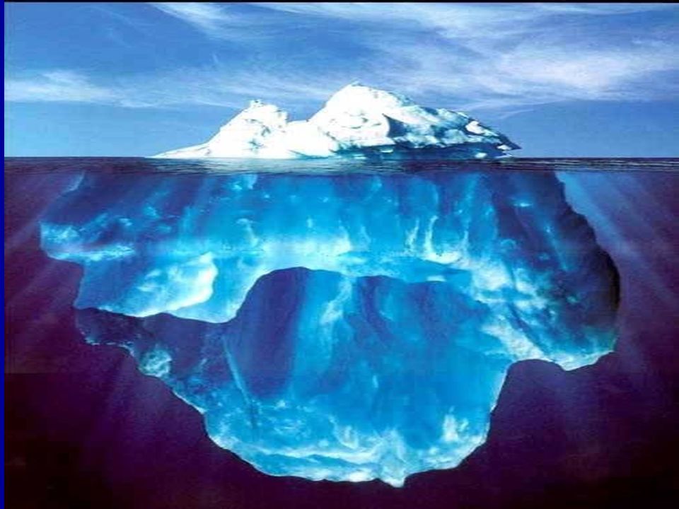
Chromosomal changes in the genome of cancer cells: tip of the iceberg
Reciprocal translocation Ring Chromosome Deletion Duplication Terminal Deletion Insertion Inversion Robertsonian Translocation Isochromosomes

19
Nucleotide changes in the genome of cancer cells: unseen site of the iceberg
Deletions Nucleotide Insertions Nucleotide Substitutions

20
DNA Loss in cancer cells

21
DNA Loss in cancer cells: beyond coincidence ...
Early Brain Tumor (Astrocytoma Stage II) Advance Brain Tumor Glioblastoma Multiform (Stage IV)
 Advance Brain Tumor. Glioblastoma Multiform (Stage IV)")
22
Mostly, it is a sign for the loss of a tumor suppressor gene
Chromosomal loss: Mostly, it is a sign for the loss of a tumor suppressor gene CDKN2 locus PTEN locus RB1 locus ??? locus p53 locus

23
Cancer: Genome Disease
Epigenetic effects

24
Genetic and Epigenetic Silencing of Tumor Suppressor Genes
Plass

25
THE CAUSES OF GENOMIC CHANGES IN CANCER
UV Replication Errors Carcinogenic chemicals Radiation Normal cell Viruses Damaged DNA Rearrangements (translocation, deletions, amplifications) Point mutations Alters DNA of genes controlling cell proliferation. (Proliferation becomes abnormal) Cancer cell
 Point mutations. Alters DNA of genes controlling cell proliferation. (Proliferation becomes abnormal) Cancer cell.")
26
Virus DNA İntegrasyonu
THE CAUSES OF GENOMIC CHANGES IN CANCER: Somatic Changes Hasar Etken Türü Etkeni Kanser Riski İşareti Fiziksel Morötesi Işınlar Deri Ka., Melanoma P53 (CC-TT) Radyasyon Tiroid Ka., Lösemi Translokasyon Kimyasal Benzopren Akciğer Ka. p53 (G-T) Aflatoksin Karaciğer Ka. p53 (249 G-T) Oksidatif Stres Yaşlılık Kanserleri P53 (C-T) Biyolojik HBV Virus DNA İntegrasyonu
 Radyasyon. Tiroid Ka., Lösemi. Translokasyon. Kimyasal. Benzopren. Akciğer Ka. p53 (G-T) Aflatoksin. Karaciğer Ka. p53 (249 G-T) Oksidatif Stres. Yaşlılık Kanserleri. P53 (C-T) Biyolojik. HBV. Virus DNA İntegrasyonu.")
27
THE CAUSES OF GENOMIC CHANGES IN CANCER: Hereditary Predisposition
Genes Disease Function Inheretance Cancer Risk FA Genes F-A DNA Damage respose ? OR Lösemi XP Genes X-P NER Type DNA Repair Skin Ca. BLM Bloom DNA Helicase ? Various cancers WRN Werner Sarcoma RECQ4 Rothmund-Thomson DNA Helicase MLH1, MSH2, PMS1, PMS2 MMR OD Colon, Endometrium Ca. Lösemi, NF1 BRCA1, BRCA2 Breast, Ovary, Prostate, Pancreas Ca ATM A-T DNA Damage sense ? Lymphoma, Leukemia Breast Ca. ? p53 Li-Fraumeni DNA Damage sense

28
CANCER AND GENETICS Approximately 90-95% of all cancers are sporadic.
5-10% are inherited.

29
GENES PLAYING ROLE IN CANCER DEVELOPMENT
• Oncogenes • Tumor suppressor genes • DNA repair genes

30
- What are the genes responsible for tumorigenic cell growth? + ++
Normal + - Proto-oncogenes Cell growth and proliferation Tumor suppressor genes Cancer Mutated or “activated” oncogenes ++ Malignant transformation Loss or mutation of Tumor suppressor genes

31
ONCOGENES Oncogenes are mutated forms of cellular proto-oncogenes.
Proto-oncogenes code for cellular proteins which regulate normal cell growth and differentiation.

32
Five types of proteins encoded by proto- oncogenes participate in control of cell growth:
Class I: Growth Factors Class II: Receptors for Growth Factors and Hormones Class III: Intracellular Signal Transducers Class IV: Nuclear Transcription Factors Class V: Cell-Cycle Control Proteins

33
Functions of Cellular Proto-Oncogenes 2. Growth Factor Receptors
1. Secreted Growth Factors 2. Growth Factor Receptors 4. Nuclear Proteins: Transcription Factors 3. Cytoplasmic Signal Transduction Proteins 5. Cell Growth Genes

34
A generic signalling pathway

35
Oncogenes proto-oncogene = ras Oncogene = mutated ras Always activated
Always stimulating proliferation

36
Amino acid substitutions in Ras family proteins (inactivates GTPase)
amino acid position Ras gene Tumor c-ras (H, K, N) Gly Ala Gln normal cells H-ras Gly Ala Leu lung carcinoma Val Ala Gln bladder carcinoma K-ras Cys Ala Gln lung carcinoma Arg Ala Gln lung carcinoma Val Ala Gln colon carcinoma N-ras Gly Ala Lys neuroblastoma Gly Ala Arg lung carcinoma Murine sarcoma virus H-ras Arg Thr Gln Harvey strain K-ras Ser Thr Gln Kirsten strain
 Gly Ala Gln normal cells. H-ras Gly Ala Leu lung carcinoma. Val Ala Gln bladder carcinoma. K-ras Cys Ala Gln lung carcinoma. Arg Ala Gln lung carcinoma. Val Ala Gln colon carcinoma. N-ras Gly Ala Lys neuroblastoma. Gly Ala Arg lung carcinoma. Murine sarcoma virus. H-ras Arg Thr Gln Harvey strain. K-ras Ser Thr Gln Kirsten strain.")
37
Activation mechanisms of proto-oncogenes
proto-oncogene --> oncogene

38
CHROMOSOMAL REARRANGEMENTS OR TRANSLOCATIONS
Neoplasm Translocation Proto-oncogene Burkitt lymphoma t(8;14) 80% of cases c-myc1 t(8;22) 15% of cases t(2;8) % of cases Chronic myelogenous t(9;22) 90-95% of cases bcr-abl2 leukemia Acute lymphocytic t(9;22) 10-15% of cases bcr-abl2 Leukemia 1c-myc is translocated to the IgG locus, which results in its activated expression 2bcr-abl fusion protein is produced, which results in a constitutively active abl kinase
 80% of cases c-myc1. t(8;22) 15% of cases. t(2;8) 5% of cases. Chronic myelogenous t(9;22) 90-95% of cases bcr-abl2. leukemia. Acute lymphocytic t(9;22) 10-15% of cases bcr-abl2. Leukemia. 1c-myc is translocated to the IgG locus, which results in its activated expression. 2bcr-abl fusion protein is produced, which results in a constitutively active abl kinase.")
39
GENE AMPLIFICATION Oncogene Amplification Source of tumor
c-myc ~20-fold leukemia and lung carcinoma N-myc ,000-fold neuroblastoma retinoblastoma L-myc fold small-cell lung cancer c-abl ~5-fold chronic myoloid leukemia c-myb fold acute myeloid leukemia colon carcinoma c-erbB ~30-fold epidermoid carcinoma K-ras fold colon carcinoma 30-60-fold adrenocortical carcinoma

40
Oncogenes are usually dominant
(gain of function) cellular proto-oncogenes that have been mutated (and “activated”) cellular proto-oncogenes that have been captured by retroviruses and have been mutated in the process (and “activated”) virus-specific genes that behave like cellular proto-oncogenes that have been mutated to oncogenes (i.e., “activated”)
 cellular proto-oncogenes that have been mutated (and activated ) cellular proto-oncogenes that have been captured by retroviruses and have been mutated in the process (and activated ) virus-specific genes that behave like cellular proto-oncogenes that have been mutated to oncogenes (i.e., activated )")
41
The result: Overproduction of growth factors
Flooding of the cell with replication signals Uncontrolled stimulation in the intermediary pathways Cell growth by elevated levels of transcription factors

42
Tumor suppressor genes
Normal function - inhibit cell proliferation Absence/inactivation of inhibitor --> cancer Both gene copies must be defective

43
KNUDSON TWO HIT HYPOTHESIS IN FAMILIAL CASES
Familial RB (%30) RB rb Normal cells rb RB rb RB LOH Inactivation of a tumor suppressor gene requires two mutations, inherited mutation and somatic mutation. Tumor cells Normal cells
 RB. rb. Normal cells. rb. RB. rb. RB. LOH. Inactivation of a tumor suppressor gene requires two mutations, inherited mutation and somatic mutation. Tumor cells. Normal cells.")
44
KNUDSON TWO HIT HYPOTHESIS IN SPORADIC CASES
RB LOH Mutation Normal Cells RB RB Inactivation of a tumor suppressor gene requires two somatic mutations. Tumor cells

45
TUMOR SUPPRESSOR GENES
Disorders in which gene is affected Gene (locus) Function Familial Sporadic DCC (18q) cell surface unknown colorectal interactions cancer WT1 (11p) transcription Wilm’s tumor lung cancer Rb1 (13q) transcription retinoblastoma small-cell lung carcinoma p53 (17p) transcription Li-Fraumeni breast, colon, syndrome & lung cancer BRCA1(17q) transcriptional breast cancer breast/ovarian tumors BRCA2 (13q) regulator/DNA repair
 Function Familial Sporadic. DCC (18q) cell surface unknown colorectal. interactions cancer. WT1 (11p) transcription Wilm’s tumor lung cancer. Rb1 (13q) transcription retinoblastoma small-cell lung. carcinoma. p53 (17p) transcription Li-Fraumeni breast, colon, syndrome & lung cancer. BRCA1(17q) transcriptional breast cancer breast/ovarian. tumors. BRCA2 (13q) regulator/DNA repair.")
46
CELL CYCLE S CELL CYCLE Daugther cell Gateway Mitosis Growth Factors
DNA replication Control Point Gateway Growth Factors Cell cycle inhibitors CELL CYCLE S

47
Rb gene Rb protein controls cell cycle moving past G1 checkpoint
Rb protein binds regulatory transcription factor E2F E2F required for synthesis of replication enzymes E2F - Rb bound = no transcription/replication Growth factor --> Ras pathway --> G1Cdk-cyclin synthesized Active G1 Cdk-cyclin kinase phosphorylates Rb Phosphorylated Rb cannot bind E2F --> S phase Disruption/deletion of Rb gene Inactivation of Rb protein --> uncontrolled cell proliferation --> cancer

48
p53 Phosphyorylated p53 activates transcription of p21 gene
p21 Cdk inhibitor (binds Cdk-cyclin complex --> inhibits kinase activity) Cell cycle arrested to allow DNA to be repaired If damage cannot be repaired --> cell death (apoptosis) Disruption/deletion of p53 gene Inactivation of p53 protein --> uncorrected DNA damage --> uncontrolled cell proliferation --> cancer
 Cell cycle arrested to allow. DNA to be repaired. If damage cannot be repaired. --> cell death (apoptosis) Disruption/deletion of p53 gene. Inactivation of p53 protein. --> uncorrected DNA damage. --> uncontrolled cell proliferation --> cancer.")
49
DNA REPAIR GENES These are genes that ensure each strand of genetic information is accurately copied during cell division of the cell cycle. Mutations in DNA repair genes lead to an increase in the frequency of mutations in other genes, such as proto-oncogenes and tumor suppressor genes. i.e. Breast cancer susceptibility genes (BRCA1 and BRCA2) Hereditary non-polyposis colon cancer susceptibility genes (MSH2, MLH1, PMS1, PMS2) have DNA repair functions. Their mutation will cause tumorigenesis.
 Hereditary non-polyposis colon cancer susceptibility genes (MSH2, MLH1, PMS1, PMS2) have DNA repair functions. Their mutation will cause tumorigenesis.")
50
Molecular mechanisms of DNA double strand break repair
BRCA1/2 Van Gent et al, 2001

51
IMPORTANCE OF DNA REPAIR

52
Tumor Progression Cellular Multiple mutations lead to colon cancer Genetic changes --> tumor changes

53
Revolution in cancer treatment: ‘Smart Bullets Period’

54
Summary of 30 years of research (1971-2001)
Hanahan & Weinberg 2000

55
Bilimsel Araştırmaların Kanserle Savaşa Katkısı
HERCEPTIN STI-571 Bilimsel Araştırmaların Kanserle Savaşa Katkısı HERCEPTİN

56
Translocation and Bcr-Abl fusion in CML

57
STI-571 against Bcr-Abl

58
Smart bullet STI-571 lockes itself to the target molecule

59
Thousands of Targets ? ? ? ? ? ? ? ? ? ? ? ? ? ? ? ? ? ? STI-571
HERCEPTIN ? ? STI-571 ? ? ? ? ? ? ?

60
MOLECULAR BIOLOGY & INFORMATICS
Biyoinformatik ~ bp DNA ~ genes ~ protein ~ interaction 1 human cell

Similar presentations


>")


