Download presentation
Presentation is loading. Please wait.

1
Chapter 10: Muscle Tissue
AnnMarie Armenti, MS SCCC BIO 130

2
Muscle Tissue A primary tissue type, divided into: skeletal muscle
Voluntary striated muscle, controlled by nerves of the central nervous system cardiac muscle Involuntary striated muscle smooth muscle Involuntary nonstriated muscle

3
Characteristics of all Muscle Tissues
Specialized Cells: - elongated, high density of myofilaments = cytoplasmic microfilaments of actin and myosin Excitability/Irritability: - receive and respond to stimulus Contractility: - shorten and produce force upon stimulation Extensibility: - can be stretched Elasticity: - recoil after stretch

4
Skeletal Muscle Tissue
Skeletal muscles make up 44% of body mass Skeletal muscle = an organ composed of: skeletal muscle cells (fibers) and CT nerves and blood vessels
 and CT. nerves and blood vessels.")
5
Functions of Skeletal Muscles
Produce skeletal movement Maintain posture and upright position Support soft tissues Guard entrances and exits Maintain body temperature by generating heat Stabilize joints

6
Muscle Tissue Organized at the Tissue Level

7
Formation of Skeletal Muscle Fibers
Skeletal muscle cells are called fibers Figure 10–2

8
Skeletal Muscle Anatomy
Each muscle is innervated by one nerve: Nerve must branch and contact each skeletal muscle fiber (cell) One artery, branches into extensive capillaries around each fiber: supply oxygen supply nutrients remove wastes.
 One artery, branches into extensive capillaries around each fiber: supply oxygen. supply nutrients. remove wastes.")
9
Organization of Connective Tissues
Figure 10–1

10
Organization of Connective Tissues
Muscles have 3 layers of connective tissues that hold the muscle together: Epimysium - covers the muscle (exterior collagen layer), separates muscle from other tissues, composed of collagen, connects to deep fascia Perimysium - composed of collagen and elastin, has associated blood vessels and nerves, bundles muscle fibers into groups called fascicles - perimysium covers a fascicle Endomysium - composed of reticular fibers, contains capillaries, nerve fibers and satellite cells (= stem cells repair), surrounds individual muscle fibers
, separates muscle from other tissues, composed of collagen, connects to deep fascia. Perimysium. - composed of collagen and elastin, has associated blood vessels and nerves, bundles muscle fibers into groups called fascicles. - perimysium covers a fascicle. Endomysium. - composed of reticular fibers, contains capillaries, nerve fibers and satellite cells (= stem cells repair), surrounds individual muscle fibers.")
11
Muscle Attachments Endomysium, perimysium, and epimysium come together: at ends of muscles to form connective tissue attachment to bone matrix Tendon = cord-like bundles Aponeurosis = sheet-like

12
How would severing the tendon attached to a muscle affect the muscle’s ability to move a body part?
Uncontrolled movement would result from a severed tendon. Movement would be greatly exaggerated with no tendon. No movement is possible without a muscle to bone connection. Limited movement would result.

13
Muscle

14
Skeletal Muscle Fibers
Huge cells: up to 100 µm diameter, 30 cm long Multinucleate Formed by fusion of 100s of myoblasts Nuclei of each myoblast retained to provide enough mRNA for protein synthesis in large fiber Unfused myoblasts in adult = satellite cells Satellite cells are capable of division and fusion to existing fibers for repair but cannot generate new fibers

15
Organization of Skeletal Muscle Fibers
Figure 10–3

16
Skeletal Muscle Fibers
Cell membrane = sarcolemma Sarcolemma maintains separation of electrical charges resulting in a transmembrane potential Na+ pumped out of the cell creating positive charge on the outside of the membrane Negative charge from proteins on inside give muscle fibers a resting potential of -85mV If permeability of the membrane is altered, Na+ will flow in causing a change in membrane potential Change in potential will signal the muscle to contract

17
Transverse Tubules Tubes of sarcolemma called transverse tubules (T tubules) reach deep inside the cell to transmit changes in transmembrane potential to structures inside the cell Transmit action potential through cell Allow entire muscle fiber to contract simulataneously
 reach deep inside the cell to transmit changes in transmembrane potential to structures inside the cell. Transmit action potential through cell. Allow entire muscle fiber to contract simulataneously.")
18
Skeletal Muscle Fibers
Cytoplasm = sarcoplasm: rich in glycosomes (glycogen granules) and myoglobin (binds oxygen) Fiber is filled with myofibrils extending the whole length of the cell Myofibrils consist of bundles of myofilaments Myofilaments are responsible for muscle contraction made of actin and myosin proteins 80% of cell volume
 and myoglobin (binds oxygen) Fiber is filled with myofibrils extending the whole length of the cell. Myofibrils consist of bundles of myofilaments. Myofilaments are responsible for muscle contraction. made of actin and myosin proteins. 80% of cell volume.")
19
Organization of Skeletal Muscle Fibers
Figure 10–3

20
Skeletal Muscle Fibers
Actin: makes up the thin filament Myosin: makes up the thick filament When thick and thin filaments interact, contraction occurs

21
Skeletal Muscle Fibers
Sarcoplasm contains networks of SER called sarcoplasmic reticulum (SR) Sarcoplasmic Reticulum: A membranous structure surrounding each myofibril Function: store calcium and help transmit action potential to myofibril SR forms chambers (terminal cisternae) attached to T-tubules Cisternae Concentrate Ca2+ (via ion pumps) Release Ca2+ into sarcomeres to begin muscle contraction All calcium is actively pumped from sarcoplasm to SR (SR has 1000X more Ca2+ than sarcoplasm)
 Sarcoplasmic Reticulum: A membranous structure surrounding each myofibril. Function: store calcium and help transmit action potential to myofibril. SR forms chambers (terminal cisternae) attached to. T-tubules. Cisternae. Concentrate Ca2+ (via ion pumps) Release Ca2+ into sarcomeres to begin muscle contraction. All calcium is actively pumped from sarcoplasm to SR (SR has 1000X more Ca2+ than sarcoplasm)")
22
Skeletal Muscle Fibers
Triads are located repeated along the length of myofilaments Triads = T-tubule wrapped around a myofibril sandwiched between two terminal cisternae of SR Formed by 1 T tubule and 2 terminal cisternae of SR Triads are located on both ends of a sarcomere Sarcomere = smallest functional unit of a myofibril

23
Sarcomere

24
Each muscle = ~ 100 fascicles
Each fascicle = ~ 100 muscle fibers Each fiber (cell) = ~ 1 thousand myofibrils Each myofibril = ~ 10 thousand sarcomeres
 = ~ 1 thousand. myofibrils. Each myofibril = ~ 10 thousand. sarcomeres.")
25
The structural components of a sarcomere.

26
Sarcomeres The contractile units of muscle
Structural units of myofibrils Form visible patterns within myofibrils

27
Sarcomeres Composed of: 1. Thick filaments – myosin
2. Thin filaments – actin 3. Stabilizing proteins: -hold thick and thin filaments in place 4. Regulatory proteins: - control interactions of thick and thin filaments Organization of the proteins in sarcomere causes striated appearance of the muscle fiber Figure 10–4

28
Muscle Striations A striped or striated pattern within myofibrils:
alternating dark, thick filaments (A bands) and light, thin filaments (I bands)
 and light, thin filaments (I bands)")
29
Regions of the Sarcomere
A-band: - whole width of thick filaments, looks dark microscopically M line: at midline of sarcomere - Center of each thick filament, middle of A-band - Attaches neighboring thick filaments H-zone: - Light region on either side of the M line - Contains thick filaments only Zone of overlap: - ends of A-bands - place where thin filaments intercalate between thick filaments (triads encircle zones of overlap)
")
30
Regions of the Sarcomere
I-band: - Contains thin filaments outside zone of overlap - Not whole width of thin filaments Z lines/disc: - the centers of the I bands - constructed of Actinins - Anchor thin filaments and bind neighboring sarcomeres - Constructed of Titin Proteins - Bind thick filaments to Z-line, stabilize the filament

32
Why does skeletal muscle appear striated when viewed through a microscope?
Z lines and myosin filaments align within the tissue. Glycogen reserves are linearly arranged. Capillaries regularly intersect the myofibers. Actin filaments repel stain, appearing banded.

33
Sarcomere Function Muscle Contraction
Transverse tubules encircle the sarcomere near zones of overlap Ca2+ released by SR causes thin and thick filaments to interact Muscle Contraction Is caused by interactions of thick and thin filaments Structures of protein molecules determine interactions

34
Thin Filament Figure 10–7a

35
Thin Filaments (5-6 nm diameter)
Made of 4 proteins: Actin Nebulin Holds F actin strands together F-actin (filamentous) consists of rows of G-actin (globular) Each G-actin has an active site that can bind to myosin Tropomyosin - Covers the active sites on G actin to prevent actin–myosin binding Troponin: holds tropomyosin on the G-actin Also has receptor for Ca2+: when Ca2+ binds to the troponin-tropomyosin complex it causes the release of actin allowing it to bind to myosin
 consists of rows of G-actin (globular) Each G-actin has an active site that can bind to myosin. Tropomyosin. - Covers the active sites on G actin to prevent actin–myosin binding. Troponin: holds tropomyosin on the G-actin. Also has receptor for Ca2+: when Ca2+ binds to the troponin-tropomyosin complex it causes the release of actin allowing it to bind to myosin.")
36
Troponin and Tropomyosin
Figure 10–7b

37
Initiating Contraction
Ca2+ binds to receptor on troponin molecule Troponin–tropomyosin complex changes Exposes active site of F actin

38
Thick Filament Figure 10–7c

39
Thick Filaments (10-12 nm diameter)
Composed of: bundled myosin molecules titin strands that recoil after stretching Each Myosin has three parts 1. Tail: - tails bundled together to make length of thick filament - all point toward M-line 2. Hinge: - flexible region, allows movement for contraction

40
Thick Filaments (10-12 nm diameter)
3. Head: - hangs off tail by hinge, will bind actin at active site. - No heads in H-zone - also contains core of titin: - elastic protein that attaches thick filaments to Z-line - Titin holds thick filament in place and aid elastic recoil of muscle after stretching - Each thick filament is surrounded by a hexagonal arrangement of thin filaments with which it will interact

41
The Myosin Molecule Figure 10–7d

42
Myosin Action During contraction, myosin heads:
interact with actin filaments, forming cross-bridges pivot, producing motion

43
Sliding Filaments Figure 10–8

44
Sliding Filament Theory
Contraction of skeletal muscle is due to thick filaments and thin filament sliding past each other not compression of the filaments H-zones and I-bands decrease width during contraction Zones of overlap increase width Z-lines move closer together A-band remains constant Sliding causes shortening of every sarcomere in every myofibril in every fiber Overall result = shortening of whole skeletal muscle

45
The components of the neuromuscular junction, and the events involved in the neural control of skeletal muscles.

46
Skeletal Muscle Contraction
Excitation Excitation-Contraction Coupling Contraction Relaxation Figure 10–9 (Navigator)
")
47
Excitation and the Neuromuscular Junction
Excitation of muscle fiber is controlled by the nervous system at the neuromuscular junction using neurotransmitter

48
The Neuromuscular Junction
Is the location of neural stimulation Action potential (electrical signal): travels along nerve axon ends at synaptic terminal
: travels along nerve axon. ends at synaptic terminal.")
49
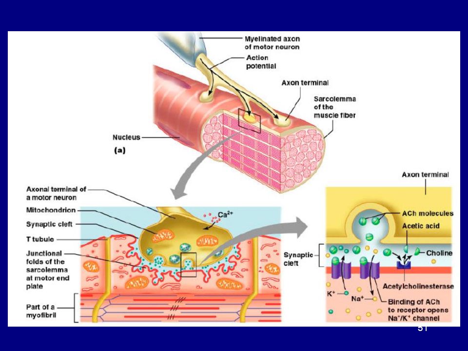
Components of Neuromuscular Junction
- where a nerve terminal interfaces with a muscle fiber at the motor end plate - one junction per fiber: control of fiber from one neuron Synaptic Terminal: - expanded end of the axon, contains vesicles of neurotransmitters Acetylcholine (Ach) Motor End Plate: - specialized sarcolemma that contains Ach receptors and the enzyme acetylcholinesterase (AchE) Synaptic Cleft: - space between the synaptic terminal and motor end plate where neurotransmitters are released
 Motor End Plate: - specialized sarcolemma that contains Ach receptors. and the enzyme acetylcholinesterase (AchE) Synaptic Cleft: - space between the synaptic terminal and motor end. plate where neurotransmitters are released.")
50
Skeletal Muscle: Neuromuscular Junction
Figure 10–10a, b (Navigator)
")
52
2. Skeletal Muscle Excitation
Figure 10–10c

53
The Neurotransmitter Acetylcholine or ACh:
travels across the synaptic cleft binds to membrane receptors on sarcolemma (motor end plate) causes sodium–ion rush into sarcoplasm is quickly broken down by enzyme (acetylcholinesterase or AChE)
 causes sodium–ion rush into sarcoplasm. is quickly broken down by enzyme (acetylcholinesterase or AChE)")
54
Action Potential Generated by increase in sodium ions in sarcolemma
Travels along the T tubules Leads to excitation–contraction coupling

55
The Process of Contraction
Neural stimulation of sarcolemma: causes excitation–contraction coupling Cisternae of SR release Ca2+: which triggers interaction of thick and thin filaments consuming ATP and producing tension

56
3. Excitation–Contraction Coupling
Action potential reaches a triad: releasing Ca2+ triggering contraction Requires myosin heads to be in “cocked” position: loaded by ATP energy

57
The key steps involved in the contraction of a skeletal muscle fiber.

58
Exposing the Active Site
The action potential of the transverse tubules reaches a triad and causes the release of calcium ions from the cisternae of the SR into the sarcoplasm around the zones of overlap of the sarcomeres Calcium binds to troponin on the thin filaments Troponin pulls tropomyosin off the active sites of the actin so that cross bridges can form. Figure 10–11

59
The Contraction Cycle Figure 10–12 (1 of 4)
")
60
5 Steps of the Contraction Cycle
Exposure of active sites Formation of cross-bridges Pivoting of myosin heads Detachment of cross-bridges Reactivation of myosin

61
The Contraction Cycle 1. Actin, free of tropomyosin, binds to myosin via its active site 2. Cross bridges are formed * Actin active sites are bound to myosin heads Figure 10–12 (2 of 4)
")
62
The Contraction Cycle Myosin heads have been pre-primed for movement via ATP energy prior to cross bridge formation and are pointed away from the M line. Upon actin binding, the myosin heads pivot toward the M line in an event called the power stroke, which pulls the thick filament along the thin filament Figure 10–12 (3 of 4)
")
63
The Contraction Cycle Myosin ATPase uses ATP to break the cross bridges releasing the myosin head from the actin active site, and resets the myosin head pointed away from the M-line

64
The Contraction Cycle The myosin head is now primed to interact with a new active site on actin Myosin can carry out 5 power strokes per second while calcium and ATP are available. Each power stroke shortens the sarcomere by 1% Figure 10–12 (Navigator) (4 of 4)
 (4 of 4)")
65
Fiber Shortening As sarcomeres shorten, muscle pulls together, producing tension Figure 10–13

66
Contraction Duration Depends on: duration of neural stimulus
number of free calcium ions in sarcoplasm availability of ATP

67
4. Relaxation Ca2+ reabsorbed by sarcoplasmic reticulum
Ca2+ ions detach from troponin Troponin, without Ca2+, pivots tropomyosin back onto active sites on actin, no cross bridges can form Sarcomeres stretch back out: Gravity Opposing muscle contractions Elastic recoil of titin protein Result: Muscle returns to Resting Length

68
A Review of Muscle Contraction
Table 10–1 (1 of 2)
")
69
A Review of Muscle Contraction
Table 10–1 (2 of 2)
")
70
Rigor Mortis A fixed muscular contraction after death Caused when:
SR can not absorb Ca2+ : ion pumps cease to function calcium builds up in the sarcoplasm Ca2+ bind troponin Tropomyosin frees actin Cross bridges from No ATP to detach myosin head because ATP is already all used up fixed cross bridge Contractions occur until necrosis releases lysosomal enzymes which digest cross bridges

71
Disease of Muscle Contraction
Botulism/Botox: Bacteria Clostridium botulinum (grows in improperly canned foods) produces botulinum toxin Toxin prevents the release of Ach at the neuromuscular junction Results in flaccid paralysis Tetanus: Bacteria Clostridium tetani (grows in soil) produces tenanus toxin: Toxin causes over stimulation of motor neurons Results in spastic paralysis Myasthenia gravis: Autoimmune disease Causes loss of Ach receptors muscles become non-responsive
 produces botulinum toxin. Toxin prevents the release of Ach at the neuromuscular junction. Results in flaccid paralysis. Tetanus: Bacteria Clostridium tetani (grows in soil) produces tenanus toxin: Toxin causes over stimulation of motor neurons. Results in spastic paralysis. Myasthenia gravis: Autoimmune disease. Causes loss of Ach receptors muscles become. non-responsive.")
72
KEY CONCEPT Skeletal muscle fibers shorten as thin filaments slide between thick filaments Free Ca2+ in the sarcoplasm triggers contraction SR releases Ca2+ when a motor neuron stimulates the muscle fiber Contraction is an active process Relaxation and return to resting length is passive

73
Where would you expect the greatest concentration of Ca2+ in resting skeletal muscle to be?
T tubules surrounding the mitochondria within sarcomeres cisternae of the sarcoplasmic reticulum

74
How would a drug that interferes with cross-bridge formation affect muscle contraction?
interferes with contraction slows contraction speeds contraction increases strength of contraction

75
Predict what would happen to a muscle if the motor end plate failed to produce acetylcholinesterase.
Muscle would lose strength. Muscle would be unable to contract. Muscle would lock in a state of contraction. Muscle would contract repeatedly.

76
What would you expect to happen to a resting skeletal muscle if the sarcolemma suddenly became very permeable to Ca2+? increased strength of contraction decreased cross bridge formation decreased ability to relax both A and C

77
The mechanism responsible for tension production in a muscle fiber, and the factors that determine the peak tension developed during a contraction.

78
Tension Production Muscle tension:
Force exerted by contracting muscle Force is applied to a load Load = weight of the object being acted upon For a single muscle fiber contraction is all–or–none: as a whole, a muscle fiber is either contracted or relaxed

79
Tension of a Single Muscle Fiber
Once contracting tension depends on: 1. The number of pivoting cross-bridges The fiber’s resting length at the time of stimulation 3. The frequency of stimulation

80
Resting Length Greatest tension produced at optimal resting length
Optimal resting length = Optimum overlap Overlap determines the number of pivoting cross-bridges Enough overlap, so that myosin can bind actin, not so much that thick filaments crash into Z-lines Figure 10–14

81
Why is it difficult to contract a muscle that has been overstretched?
Myosin filaments break. Crossbridges can not be formed. Z lines are unable to sustain contractile forces. Tendons lose elasticity.

82
Frequency of Stimulation
Twitch = single contraction due to a single neural stimulation, 3 phases: Latent period: post stimulation but no tension Action potential moves across the sarcolemma Ca2+ is released Contraction phase: peak tension production - Ca2+ bind - Active cross bridge formation Relaxation phase: decline in tension Ca2+ is reabsorbed Cross bridges decline

83
Myogram A graph of twitch tension development

84
Twitch Single twitch will not produce normal movement
requires many cumulative twitches Repeat stimulation will result in higher tension due to Ca2+ not being fully absorbed - Ca2+ more cross bridges Types of Frequency Stimulation Treppe Wave summation Incomplete Tetanus Complete Tetanus

85
Treppe Stepping up of tension production to max level with repeat stimulation of the same fiber following relaxation phase Repeated stimulations immediately after relaxation phase: stimulus frequency < 50/second Causes a series of contractions with increasing tension

86
Treppe A stair-step increase in twitch tension Figure 10–16a

87
Wave Summation Repeat stimulation before relaxation phase ends resulting in more tension production than max treppe stimulus frequency > 50/second Typical muscle contraction Increasing tension or summation of twitches Figure 10–16b

88
Incomplete Tetanus Rapid cycles of contraction and relaxation produces max tension Twitches reach maximum tension Cardiac muscle incomplete tetanus Only to prevent seizure of heart Figure 10–16c

89
Complete Tetanus Relaxation eliminated, continuous contraction
Fiber is in prolonged state of contraction Produces 4x more tension than maximum treppe Quick to fatigue Most Skeletal muscle complete tetanus when contracting Figure 10–16d

90
Increased blood flow improves contraction.
During treppe, why does tension in a muscle gradually increase even though the strength and frequency of the stimulus are constant? Increased blood flow improves contraction. Sarcomeres shorten with each contraction. Calcium ion concentration increases with successive stimuli. Generated heat improves contraction.

91
The factors that affect peak tension production during the contraction of an entire skeletal muscle, and the significance of the motor unit in this process.

92
Tension Produced by Whole Skeletal Muscles
Depends on: Internal tension produced by sarcomeres - Not all the tension is transferred to the load, some of it is lost due to the elasticity of muscle tissues External tension exerted by muscle fibers on elastic extracellular fibers - Tension applied to the load 3. Total number of muscle fibers stimulated

93
Total Number of Muscle Fibers Stimulated
Each skeletal muscle has thousands of fibers organized into motor units Motor units = all fibers controlled by a single motor neuron Axon branches to contact each fiber Number of fibers in a motor unit depends on the function Fine control: 4/unit (e.g. eye muscles) Gross control: 2000/unit (e.g. leg muscles) Fibers from different units are intermingled in the muscle so that the activation of one unit will produce equal tension across the whole muscle
 Gross control: 2000/unit (e.g. leg muscles) Fibers from different units are intermingled in the muscle so that the activation of one unit will produce equal tension across the whole muscle.")
94
Motor Units in a Skeletal Muscle
Figure 10–17

95
Recruitment (Multiple Motor Unit Summation)
In a whole muscle or group of muscles, smooth motion and increasing tension is produced by slowly increasing size or number of motor units stimulated Recruitment = order of activation of a motor unit Slower weaker units are activated first Strong units are added to produce steady increases in tension

96
Contraction Skeletal Muscle
During sustained contraction of a muscle Some units rest while others contract to avoid fatigue For maximum tension, all units in complete tetanus Leads to rapid fatigue Muscle tone = maintaining shape/definition of the muscle Some units are always contracting Exercise = Increase # of units contraction Increase in metabolic rate Increase in speed of recruitment (better tone)
")
97
KEY CONCEPT Voluntary muscle contractions involve sustained, tetanic contractions of skeletal muscle fibers Force is increased by increasing the number of stimulated motor units (recruitment)
")
98
The types of muscle contractions.

99
Contraction Skeletal Muscle
All contractions produce tension but not always movement Isotonic Contractions: - Muscle length changes resulting in movement Isometric Contractions - Tension is produced with no movement

100
Isotonic Contraction If muscle tension > resistance:
muscle shortens (concentric contraction) If muscle tension < resistance: muscle lengthens (eccentric contraction) Figure 10–18a, b
 If muscle tension < resistance: muscle lengthens (eccentric contraction) Figure 10–18a, b.")
101
Isometric Contraction
Skeletal muscle develops tension, but is prevented from changing length Note: Iso = same, metric = measure Figure 10–18c, d

102
Return to Resting Length
Expansion via: Elastic recoil after contraction The pull of elastic elements (tendons and ligaments) Expands the sarcomeres to resting length Opposing muscle contractions - Reverse the direction of the original motion Gravity - Opposes muscle contraction to return a muscle to its resting state
 Expands the sarcomeres to resting length. Opposing muscle contractions. - Reverse the direction of the original motion. Gravity. - Opposes muscle contraction to return a muscle to its resting state.")
103
Can a skeletal muscle contract without shortening? Explain.
Yes; isotonic contractions produce no movement. No; resistance is always less than force generated. Yes; concentric contractions are common. No; contraction implies movement.

104
The mechanisms by which muscle fibers obtain energy to power contractions.

105
Muscle Metabolism 1 fiber ~15 million thick filaments
1 thick filament ~ 2500 ATP/sec 1 glucose (aerobic respiration) = 36 ATP Each fiber needs 1x10¹² glucose/sec to contract ATP unstable, muscles store respiration energy on creatine as Creatine Phosphate (CP) Creatine phosphokinase transfers P from CP at ADP when ATP is needed to reset myosin for next contraction Each cell as only ~20 sec of energy reserved
 = 36 ATP. Each fiber needs 1x10¹² glucose/sec to contract. ATP unstable, muscles store respiration energy on creatine as Creatine Phosphate (CP) Creatine phosphokinase transfers P from CP at ADP when ATP is needed to reset myosin for next contraction. Each cell as only ~20 sec of energy reserved.")
106
ATP and CP Adenosine triphosphate (ATP): Creatine phosphate (CP):
the active energy molecule Creatine phosphate (CP): the storage molecule for excess ATP energy in resting muscle Energy recharges ADP to ATP: using the enzyme creatine phosphokinase (CPK) When CP is used up, other mechanisms generate ATP
: the storage molecule for excess ATP energy in resting muscle. Energy recharges ADP to ATP: using the enzyme creatine phosphokinase (CPK) When CP is used up, other mechanisms generate ATP.")
107
Muscle Metabolism At Rest: Moderate Activity:
Use glucose and fatty acids with O2 (from blood) aerobic respiration Resulting ATP is used to CP reserves Excess glucose is stored as glycogen Moderate Activity: CP used up Glucose and fatty acids with O2 (from blood) are used to generate ATP (aerobic respiration)
 aerobic respiration. Resulting ATP is used to CP reserves. Excess glucose is stored as glycogen. Moderate Activity: CP used up. Glucose and fatty acids with O2 (from blood) are used to generate ATP (aerobic respiration)")
108
Muscle Metabolism High Activity: O2 not delivered adequately
Glucose from glycogen reserves are used for ATP via fermentation (glycolysis only) Pyruvic acid is converted to lactic acid Excess lactic acid production leads to muscle cramps
 Pyruvic acid is converted to lactic acid. Excess lactic acid production leads to muscle cramps.")
109
ATP Generation Cells produce ATP in 2 ways:
aerobic metabolism of fatty acids in the mitochondria (At rest and Moderate activity) Is the primary energy source of resting muscles Breaks down fatty acids Produces 34 ATP molecules per glucose molecule anaerobic glycolysis (fermentation) in the cytoplasm (High activity) Is the primary energy source for peak muscular activity Produces 2 ATP molecules per molecule of glucose Breaks down glucose from glycogen stored in skeletal muscles
 Is the primary energy source of resting muscles. Breaks down fatty acids. Produces 34 ATP molecules per glucose molecule. anaerobic glycolysis (fermentation) in the cytoplasm (High activity) Is the primary energy source for peak muscular activity. Produces 2 ATP molecules per molecule of glucose. Breaks down glucose from glycogen stored in skeletal muscles.")
110
Muscle Metabolism Figure 10–20a

111
Muscle Metabolism Figure 10–20b

112
Muscle Metabolism Figure 10–20c

113
Muscle Metabolism Figure 10–20 (Navigator)
")
114
Factors that contribute to muscle fatigue, and the stages and mechanisms involved in muscle recovery.

115
Muscle Fatigue When muscles can no longer perform a required activity (contraction), they are fatigued Depletion of reserves - glycogen, ATP, CP Decreased pH due to: lactic acid production Damage to sarcolemma and sarcoplasmic reticulum Muscle exhaustion and pain

116
To restore function, cell need:
Intracellular energy reserves - Glycogen and CP Good Circulation - Nutrients in, wastes out Normal O2 levels Normal pH Lactic Acid Disposal

117
Normal pH Lactic Acid Disposal
Lactic acid diffuses into the blood Filtered out by the liver Converted back to glucose through the Cori Cycle Returned to blood for use by cells When O2 returns Remaining lactic acid in the muscle is converted to glucose and used in aerobic cellular respiration

118
KEY CONCEPT Skeletal muscles at rest metabolize fatty acids and store glycogen During light activity, muscles generate ATP through aerobic breakdown of carbohydrates, lipids or amino acids At peak activity, energy is provided by anaerobic reactions that generate lactic acid as a byproduct

119
Muscle fibers and physical conditioning that relate to muscle performance.

120
Muscle Performance Power: Endurance: Power and endurance depend on:
the maximum amount of tension produced Endurance: the amount of time an activity can be sustained Power and endurance depend on: Types of muscle fibers Fast Glycolytic Fibers (fast twitch) Slow Oxidative Fibers (slow twitch) Intermediate/Fast Oxidative Fibers Physical conditioning Aerobic Exercise Resistance Exercise
 Slow Oxidative Fibers (slow twitch) Intermediate/Fast Oxidative Fibers. Physical conditioning. Aerobic Exercise. Resistance Exercise.")
121
Fiber Types Types of fibers in a muscle are genetically determined and mixed Fast glycolytic Fibers (fast twitch) Myosin ATPase work quickly Anaerobic ATP production: glycolysis only Large diameter fibers More myofilaments and glycogen Few mitochondria Fast to act, powerful, but quick to fatigue Catabolize glucose only

122
Fiber Types Slow Oxidative Fibers (slow twitch)
Myosin ATPases work slowly Specialized for aerobic respiration Many mitochondria Extensive blood supply Myoglobin (red pigment, binds oxygen) Smaller fibers for better diffusion Slow to contract, weaker tension, but resist fatigue Catabolize glucose, lipids, and amino acids
 Smaller fibers for better diffusion. Slow to contract, weaker tension, but resist fatigue. Catabolize glucose, lipids, and amino acids.")
123
Fiber Types 3. Intermediate/Fast Oxidative Fibers
Qualities of both fast glycolytic and slow oxidative fibers Fast acting but perform aerobic respiration so to resist fatigue Physical conditioning can convert some fast fibers into intermediate fibers for stamina

124
Fast versus Slow Fibers
Figure 10–21

125
Comparing Skeletal Muscle Fibers
Table 10–3

126
Muscles and Fiber Types
White muscle: mostly fast fibers pale (e.g., chicken breast) Red muscle: mostly slow fibers dark (e.g., chicken legs) Most human muscles: mixed fibers pink
 Red muscle: mostly slow fibers. dark (e.g., chicken legs) Most human muscles: mixed fibers. pink.")
127
Physical Conditioning
Aerobic Exercise: - Increase Capillary Density Increase Mitochondria and myoglobin Both then: Increase efficiency of muscle metabolism Increase strength and stamina Decrease fatigue Resistance Exercise: Results in Hypertrophy: fibers increase in diameter but not number Increase glycogen, myofibrils, and myofilaments results in increase tension production

128
Physical Conditioning
Growth Hormone (pituitary) and Testosterone (male sex hormone) Stimulate synthesis of contractile proteins Results in Muscle Enlargement Epinephrine Stimulates increase muscle metabolism Results in increase force of contraction Without stimulation muscles will atrophy Fibers shrink due to loss of myofilament proteins Loss: up to ~5%/day
 and Testosterone (male sex hormone) Stimulate synthesis of contractile proteins. Results in Muscle Enlargement. Epinephrine. Stimulates increase muscle metabolism. Results in increase force of contraction. Without stimulation muscles will atrophy. Fibers shrink due to loss of myofilament proteins. Loss: up to ~5%/day.")
129
KEY CONCEPT What you don’t use, you loose
Muscle tone indicates base activity in motor units of skeletal muscles Muscles become flaccid when inactive for days or weeks Muscle fibers break down proteins, become smaller and weaker With prolonged inactivity, fibrous tissue may replace muscle fibers

130
Why would a sprinter experience muscle fatigue before a marathon runner would?
Sprinters cannot utilize ATP for long periods of time. Sprinters’ muscles are most efficient aerobically. Sprinters’ muscles are most efficient anaerobically. Sprinters’ muscles are weaker.

131
Which activity would be more likely to create an oxygen debt: swimming laps or lifting weights?
both A and B neither A nor B

132
thick, glycogen-laden fibers
Which type of muscle fibers would you expect to predominate in the large leg muscles of someone who excels at endurance activities, such as cycling or long-distance running? slow fibers fast fibers nonvascular fibers thick, glycogen-laden fibers

133
Cardiac Muscle Tissue

134
Cardiac Muscle Tissue Cardiac muscle is striated, found only in the heart Figure 10–22

135
Cardiac Muscle Tissue Forms the majority of heart tissue
Cells = cardiocytes One or two nuclei No cell division Long branched cells Myofibrils organized into sarcomeres (striated) No triads (no terminal cisternae) Transverse tubules encircle Z-lines Aerobic Respiration Only Mitochondria and myoglobin rich Glycogen and lipid energy reserves Intercalated discs at cell junctions (gap junctions and desmosomes) allow transmission of action potentials link myofibrils from on cardiocyte (cell) to the next
 No triads (no terminal cisternae) Transverse tubules encircle Z-lines. Aerobic Respiration Only. Mitochondria and myoglobin rich. Glycogen and lipid energy reserves. Intercalated discs at cell junctions (gap junctions and desmosomes) allow transmission of action potentials. link myofibrils from on cardiocyte (cell) to the next.")
136
Coordination of Cardiocytes
Because intercalated discs link heart cells mechanically, chemically, and electrically, the heart functions like a single, fused mass of cells

137
4 Functions of Cardiac Tissue
Automaticity: contraction without neural stimulation Automatically due to control by pacemaker cells These cells generate action potentials spontaneously Pace and amount of contraction tension: Can be adjusted and controlled by the nervous system Extended contraction time - Contraction is 10x longer than skeletal muscle Only twitches, no complete tetanus - Prevention of wave summation and tetanic contractions by cell membranes

138
Smooth Muscle Tissue

139
Structure of Smooth Muscle
Nonstriated tissue Figure 10–23

140
Smooth Muscle Tissue Lines hollow organs Forms errector pili muscles
Regulates blood flow and movement of materials in organs Forms errector pili muscles Usually organized into two layer Circular Longitudinal Spindle shaped cells Single central nucleus Cells capable of division No myofibrils, sarcomeres, or T tubules SER/ER throughout cytoplasm No tendons

141
Smooth Muscle Tissue Thick filaments (myosin fibers) scattered
Myosin fibers have more heads per thick filament Thin filaments are attached to dense bodies on desmin cytoskeleton (web) Adjacent cells attach at dense bodies with gap junctions (firm linkage and communication) Dense bodies transmit contractions from cell to cell Contraction compresses the whole cell
 Adjacent cells attach at dense bodies with gap junctions (firm linkage and communication) Dense bodies transmit contractions from cell to cell. Contraction compresses the whole cell.")
142
Smooth Muscle in Body Systems
Forms around other tissues In blood vessels: regulates blood pressure and flow In reproductive and glandular systems: produces movements In digestive and urinary systems: forms sphincters produces contractions In integumentary system: arrector pili muscles cause goose bumps

143
Smooth Excitation-Contraction
Different than striated muscle: no troponin so active sites on actin are always exposed Events: Stimulation causes Ca2+ release from SR Ca2+ binds calmondulin in the sarcoplasm - Calmondulin = CALcium MODULated proteIN Calmondulin activates myosin light chain kinase, this complex phosphorylates myosin MLC Kinase converts ATP ADP to cock myosin head Cross bridge form contraction, cells pull toward center

144
Smooth Excitation-Contraction
Stimulation is by involuntary control from - Autonomic Nervous System - Hormones - Other Chemical Factors Skeletal Muscle = Motor Neurons Cardiac Muscle = Automatically

145
Characteristics of Skeletal, Cardiac, and Smooth Muscle
Table 10–4

146
Extracellular Ca2+ inhibits actin.
Why are cardiac and smooth muscle contractions more affected by changes in extracellular Ca2+ than are skeletal muscle contractions? Extracellular Ca2+ inhibits actin. Crossbridges are formed extracellularly. Most calcium for contractions comes from SR stores. Most calcium for contractions comes from extracellular fluid.

147
Smooth muscle can contract over a wider range of resting lengths than skeletal muscle can. Why?
Smooth muscle sarcomeres are longer. Myofilament arrangement is less organized in smooth muscle. Smooth muscle cells are shorter. Smooth muscle actin is longer.

148
Effects of Aging Skeletal Muscle fibers become thinner
Decrease myofibrils, Decrease reserves = Decrease in strength and endurance and Increase in fatigue Decrease cardiac and smooth muscle function = Decrease cardiovascular performance Increase fibrosis (CT): Skeletal muscle less elastic Decrease ability to repair Decrease satellite cells Increase scar formation
: Skeletal muscle less elastic. Decrease ability to repair. Decrease satellite cells. Increase scar formation.")
149
SUMMARY 3 types of muscle tissue: Functions of skeletal muscles
cardiac smooth Functions of skeletal muscles Structure of skeletal muscle cells: endomysium perimysium epimysium Functional anatomy of skeletal muscle fiber: actin and myosin

150
SUMMARY Nervous control of skeletal muscle fibers:
neuromuscular junctions action potentials Tension production in skeletal muscle fibers: twitch, treppe, tetanus Tension production by skeletal muscles: motor units and contractions Skeletal muscle activity and energy: ATP and CP aerobic and anaerobic energy

151
SUMMARY Skeletal muscle fatigue and recovery
3 types of skeletal muscle fibers: fast, slow, and intermediate Skeletal muscle performance: white and red muscles physical conditioning Structures and functions of: cardiac muscle tissue smooth muscle tissue AnnMarie Armenti, MS SCCC BIO 130

Similar presentations


 Generate heat - body temp 3 types: Skeletal - moves bone, voluntary Smooth.>")


 –sheet or band of fibrous C.T. under.>")

