Download presentation
Presentation is loading. Please wait.

1
Reproductive Emergencies
Jeanette Yamamoto, DVM

2
Canine Pregnancy – the Basics
Litter Size determination In general, the larger the breed, the larger the average litter size. Other factors: Sperm count of the male Timing of breeding and frequency of breeding Health of the bitch and condition of her uterus Bitch’s nutritional status, underlying diseases, and exposure to pharmaceuticals

3
Canine Pregnancy – the Basics
Normal gestation length (avg. 65) days from the time of first breeding
 days from the time of first breeding.")
4
Feline pregnancy – the Basics
Induced ovulators Normal gestation length – days from the time of first breeding

5
Pregnancy – the Basics Pre-parturition signs: Mammary development
Vulvar enlargement Mucoid vaginal discharge Relaxation of the pelvic ligaments Onset of lactation (24 hrs. prior to parturition) Body temp. <99 F (24 hrs. prior to parturition)
 Body temp. <99 F (24 hrs. prior to parturition)")
6
Stages of Parturition Stage 1: Typically lasts for 6 to 24 hrs.
Subclinical uterine contractions, progressive dilation of cervix Bitches: restless, panting, nesting behaviors, hiding, and anorexia. Queens: tachypneic, restless, vocal, purring, and may stay near a nesting box/area.

7
Stages of Parturition Stage 2: Active expulsion of the fetuses
First fetus delivered within 1 hr. of onset of Stage 2, with subsequent fetuses every 15 min. to 4 hrs.. Entire process occurs within 2-12 hrs., but may take as long as 24 hrs. with larger litters.

8
Stages of Parturition Stage 3: Expulsion of the placenta
Should be one placenta for each fetus delivered Placentas are usually attached to umbilical cord and will be expulsed with each birth. If placenta becomes detached, expulsion should occur several hours after birth.

9
“Green Vaginal Discharge”
Lochia = greenish vaginal discharge Indicates placental separation May be seen during ALL stages of labor and does not necessarily indicate a need for concern! Reddish-brown vaginal discharge expected for 4-6 weeks post-parturition.

10
“Guadalupe” – 2 yr. F/I Chihuahua
Presented with puppy (dead) protruding from vulva Owner stated pet had been this way for 6 hrs. No “known” medical history Vitals – HR 120; light pink, tacky MM PE – 5% dehydrated; live fetus palpated in abdomen; prominent mammary glands
 protruding from vulva. Owner stated pet had been this way for 6 hrs. No known medical history. Vitals – HR 120; light pink, tacky MM. PE – 5% dehydrated; live fetus palpated in abdomen; prominent mammary glands.")
11
Dystocia Dystocia: Difficult parturition to the point of needing human intervention. Reasons to head to the ER: No signs of labor hours after temp. <100 F Active uterine contractions >1 hr. without expulsion of fetus More than 3 (up to 4) hours between births No fetus produced 4 hours after onset of labor Failure to initiate labor at the end of gestation Abnormal vaginal discharge More than 1 week overdue
 hours between births. No fetus produced 4 hours after onset of labor. Failure to initiate labor at the end of gestation. Abnormal vaginal discharge. More than 1 week overdue.")
12
Dystocia – WHY? Maternal factors
Uterine inertia (most common) Previous pelvic fractures Reproductive tract issues: vaginal stricture, insufficient cervical dilation, vaginal mass, uterine torsion Inguinal hernia Extreme nervousness of the dam Morphologic factors – obstruction of birth canal
 Previous pelvic fractures. Reproductive tract issues: vaginal stricture, insufficient cervical dilation, vaginal mass, uterine torsion. Inguinal hernia. Extreme nervousness of the dam. Morphologic factors – obstruction of birth canal.")
13
Dystocia – WHY? Fetal factors
Malpresentation (**NOT posterior presentation) Head first with retention of forelegs One foreleg forward and retention of the other Dorsiflexion of the head Transverse presentation with fetal head in one uterine horn, rear quarters in the other. Oversized fetus Fetal malformations Fetal death
 Head first with retention of forelegs. One foreleg forward and retention of the other. Dorsiflexion of the head. Transverse presentation with fetal head in one uterine horn, rear quarters in the other. Oversized fetus. Fetal malformations. Fetal death.")
14
Dystocia - Signalment Primiparous bitches < 2 years old.
Toy and small brachycephalic breeds Very large or very small litters Not as common in cats

15
Clinical Signs Vocalization Biting/chewing at vulvar region
Abnormal vulvar discharge Fetus protruding from vulva Systemic illness – fever, weakness, tremors

16
Dystocia – PE & Diagnostics
Vaginal exam – sterile technique Abdominal radiographs Assess size, number, position and location of the fetuses /- fetal viability Assess maternal pelvic structure and status of abdomen

17
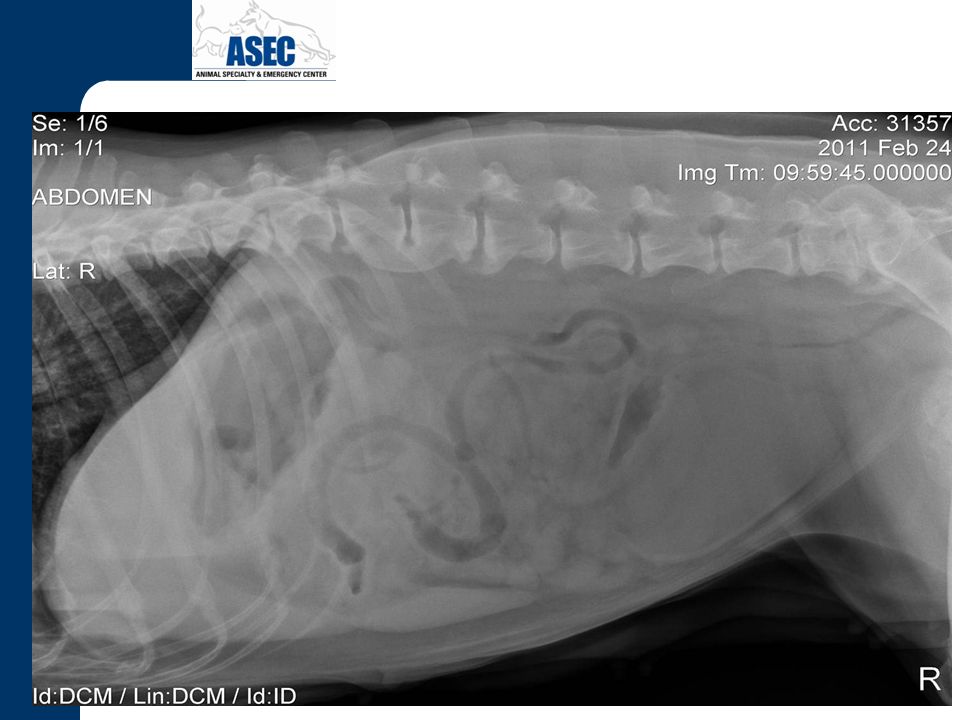
Abdominal Radiographs
Mummified fetus Normal fetal structure, size

18
Dystocia – PE & Diagnostics
Abdominal ultrasound Assess fetal viability, malformations Fetal HR: dogs bpm, cats bpm Minimum database PCV/TS, calcium level (total + ionized), BG Ideally, full CBC + chemistry panel
, BG. Ideally, full CBC + chemistry panel.")
19
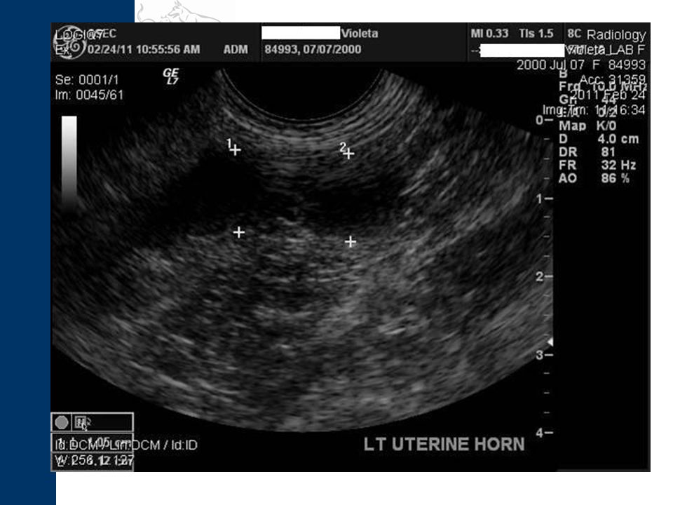
Abdominal Ultrasound Fetal head and thorax

20
Back to “Guadalupe”

21
Fetus In the Birth Canal
Attempt manual removal of fetus Sterile lube (diluted) +/- sterile red rubber catheter Dam standing or laterally recumbent + caudal abdominal palpation Posterior-ventral traction of the fetus Obstetrical instruments should be avoided An episiotomy can be performed at the dorsal vulvar midline if the vulvar opening is too narrow. If unable to remove fetus --- SURGERY!!
 +/- sterile red rubber catheter. Dam standing or laterally recumbent + caudal abdominal palpation. Posterior-ventral traction of the fetus. Obstetrical instruments should be avoided. An episiotomy can be performed at the dorsal vulvar midline if the vulvar opening is too narrow. If unable to remove fetus --- SURGERY!!")
22
Digital manipulation and extraction of fetus
12-14 g. red rubber with sterile lube via syringe

23
Possible causes for Guadalupe’s dystocia:
1. Primary uterine inertia 2. Fetus presenting head first with retention of forelegs

24
Oxytocin Treat hypoglycemia, hypocalcemia prior to use
Considerations – obstruction of birth canal, oversized fetus, abnormal presentation of fetus Uterine rupture = fetal death

25
Oxytocin Dosage Dogs: 0.5-2 units IM (or IV) – some list up to 20 u.
Cats: 0.5 units IM (or IV) – do not exceed 3 u. If no fetus, can repeat dosing 30 minutes later +/-0.5 mg/kg 10% Dextrose slowly IV. No additional doses of oxytocin recommended past this point (though some references list 3rd dosing)
 – do not exceed 3 u. If no fetus, can repeat dosing 30 minutes later +/-0.5 mg/kg 10% Dextrose slowly IV. No additional doses of oxytocin recommended past this point (though some references list 3rd dosing)")
26
Calcium Gluconate 10% Dose as a uterotonic agent: 11mg/kg diluted in saline administered IV over minutes while monitoring EKG. If noted hypocalcemia, administer mg/kg IV over min. while monitoring EKG.

27
Prognosis Guarded 20-40% success with medical management
Dystocia lasting >6hr = increased stillbirth rates Recent studies – With C-sections, neonatal survival rate reported at 92% 80% survival 7 days post C-section.

28
Off to C-Section… Anesthesia Agents Goal: survival of dam and neonates
Minimize exposure to fetuses Prep dam prior to induction Pre-oxygenate dam Agents Glycopyrrolate Propofol Gas anesthesia (+/- mask down) Regional anesthesia: line blocks, epidurals
 Regional anesthesia: line blocks, epidurals.")
29
Surgery Prophylactic antibiotics, anti-emetics?? Cesarean without OHE
+/- oxytocin OHE En bloc resection **Decision made based on dam and fetal status, staff available, and surgeon’s choice.

30
Neonate resuscitation
Suction nares, nasopharynx +/- swing technique Rubbing/drying, stimulation Acupuncture point – GV 26 (Jen Chung) Oxygen support +/- Naloxone, Doxapram, Epinephrine sublingual
 Oxygen support. +/- Naloxone, Doxapram, Epinephrine sublingual.")
31
Neonate resuscitation
Umbilical cord care Heat support KEYS: Be patient!! Listen for that sweet sound!!

32
Neonate care Once dam is recovered and relaxed, attach neonates to nipples and encourage suckling Oxytocin release

33
Success!!

34
“Violeta” – 10 yr. F/I Labrador
Inappropriate urination, defecation, for a few days. Vomited multiple times at home. Seen by rDVM - possible splenic mass on palpation & X-rays. Abdominocentesis revealed frank blood. Owner opted to take Violeta home to make decision.

35
“Violeta” Presented to ASEC 2 days later for surgical consult
Vitals: WNL PE: Small amount of sanguinopurulent vaginal discharge. No masses palpable but dorsal cranial wall of vagina feels slightly rough/irregular Firm bony swelling palpable at the pubis Abdominal radiographs….

38
Abdominal Ultrasound 1. Uterine pyometra, mucometra, etc. with concurrent endometrial thickening and/or endometritis. 2. Suspect bilateral cystic ovaries. 3. Mild bilateral renal pyelectasia may represent pyelonephritis or another nephropathy. 4. Right renal cortical cysts likely represent a degenerative change. 5. Mild left adrenomegaly may be a variation normal of represent benign hypertrophy. 6. Irregular, proliferative pubic bone contour may represent neoplasia or osteomyelitis.

41
Suspected diagnosis….. PYOMETRA!!!

42
Pyometra Secondary to cystic endometrial hyperplasia in response to endogenous progesterone Develops when bacteria invade the abnormal endometrium, leading to intraluminal accumulation of pus. Cats – Not CEH (cystic endometrial hyperplasia); ovulation by coitus, spontaneous ovulation
; ovulation by coitus, spontaneous ovulation.")
43
Pyometra Most common bacteria: Escherichia coli
Other causes: foreign body, ovarian remnant Gram negative rods

44
Signalment Older bitches Queens < 6 years old
**4 weeks to 4 months post-estrus Bitches with short interestrous intervals Young animals treated with exogenous estrogen or progestagen. +/- genetic predisposition in certain lines

45
Clinical signs Anorexia Lethargy Depression Polyuria Polydipsia
Vomiting Diarrhea +/- Febrile +/- Vaginal discharge +/- Abdominal distention **Systemic illness, shock with closed presentation

46
Diagnostics CBC: immature neutrophilia, leukopenia; mild normocytic, normochromic anemia Chemistry: Azotemia, elevated protein and globulins, +/- elevated ALT, ALP, electrolyte disturbances

47
“Violeta” In-house CBC: In-house Chemistry: No UA performed
LYMPH 8600 (H), GRAN 0, HCT 37.4% MCV 63.3 (WNL), MCHC 38.0 (WNL) PLT 187,000 (L) In-house Chemistry: ALP 207(H) Na+ 132 (L) No UA performed
, GRAN 0, HCT 37.4% MCV 63.3 (WNL), MCHC 38.0 (WNL) PLT 187,000 (L) In-house Chemistry: ALP 207(H) Na+ 132 (L) No UA performed.")
48
Diagnostics UA: **NO CYSTO!! Proteinuria +/- UTI USG:
>1.030 = pre-renal uremia <1.006 = nephrogenic diabetes insipidus secondary to E. coli. = unsure of renal function at that time and will need to monitor, assess lytes

49
Azotemia – in depth E. coli interferes with Na+ & Cl- absorption in the loop of Henle, thus reducing medullary hypertonicity and resulting in impaired water absorption. E. coli endotoxins block the action of ADH on the collecting ducts, resulting in PU/PD.

50
Diagnostics Vulvar discharge cytology: degenerative polymorphonuclear cells and bacteria; may be normal with closed cervix Abdominal Radiographs: *Don’t be fooled!! Abdominal Ultrasound: best diagnostic tool

51
Obvious Much???

53
Medical management May be initiated if….
Open pyometra Future breedings planned Not systemically ill Antibiotics: fluoroquinolones – 3-4 weeks Prostaglandin therapy – lyse corpora lutea and cause myometrial contraction.

54
Prostaglandin therapy
PGF2α dosing (off-label) Dogs: mg/kg SQ BID to TID for 3-7 days until no intrauterine fluid is noted via ultrasound Cats: mg/kg SQ BID to TID for 2-5 days until the size of the uterus is near normal Only natural prostaglandins should be used. Should be avoided in cases of closed pyometra.
 Dogs: mg/kg SQ BID to TID for 3-7 days until no intrauterine fluid is noted via ultrasound. Cats: mg/kg SQ BID to TID for 2-5 days until the size of the uterus is near normal. Only natural prostaglandins should be used. Should be avoided in cases of closed pyometra.")
55
Prostaglandin therapy
Side-effects: anxiety, vomiting, tachypnea, and tachycardia within minutes of administration. Animals should be hospitalized and monitored during initial administration. If vaginal discharge is still present 2 weeks following a five-day course of prostaglandin therapy, a second round of therapy should be initiated. The pet should be bred on the very next estrus.

56
Surgery = “Glorified Spay”
Routine OHE approach Uterus may be friable and prone to rupture Warm, sterile saline flush if fluid leakage Monitor BP, temp closely intra-op

57
“Violeta” Surgery - Both ovaries were palpably irregular textured and the left uterine horn was dilated. OHE performed. 50% debulking of pubic mass. Culture + MIC: E.coli and Bacteroides fragilis Histopath of mass: Chondrosarcoma Discharged from hospital 48 hr. post-op

58
Eclampsia a.k.a. - Puerperal tetany
Result of hypocalcemia in postpartum or peripartum animals. Occurs most commonly within the first 21 days after whelping. Small and medium breed dogs Rare in cats

59
Clinical signs Early signs: Later signs: Restlessness, pacing Panting
Whining Salivation Tremors Stiffness Tonic-clonic muscle spasms Fever Tachycardia Miosis Seizures Death

60
Calcium Gluconate 10% Administer mg/kg IV over min. while monitoring EKG. Can cause vomiting during administration. Pet will usually become more alert and muscle tremors subside.

61
Additional treatment IV catheter, IV fluids
Dextrose supplementation PRN A second dose of calcium gluconate diluted 50:50 with saline can be given subcutaneously.

62
Long term management Oral calcium treatment
Dose: 50mg/kg PO TID Should be continued throughout lactation Puppies should be weaned early Puppies should be removed and hand-raised if eclampsia occurs again.

63
Retained placenta Retained placenta within the uterus can predispose the dam to metritis. Clinical signs: Foul smelling discharge Lethargy Fever Toxemia Vomiting Death Anorexia

64
Retained placenta PE: May be able to palpate placenta on abdominal palpation. Confirm diagnosis based on abdominal ultrasound. Treatment: Antibiotics PGF2α (preferred if >24 hr. post delivery) or Oxytocin IV fluids, hospitalization if needed
 or Oxytocin. IV fluids, hospitalization if needed.")
65
Retained placenta Puppies or kittens should be allowed to nurse if the dam is not systemically affected. Be aware that if the bitch is not watched very closely she may eat the placenta before it is seen.

66
Metritis (Postpartum endometritis)
Metritis occurs in the post partum period (1-7 days later) Metritis, as opposed to pyometra, occurs when progesterone levels are low. Due to ascending infection following uterine damage from whelping, abortion, dystocia, retained placenta, retained fetus, or fetal infection.
 Metritis, as opposed to pyometra, occurs when progesterone levels are low. Due to ascending infection following uterine damage from whelping, abortion, dystocia, retained placenta, retained fetus, or fetal infection.")
67
Metritis Clinical signs: Anorexia Depression Fever Vomiting
Weight loss in puppies due to decreased lactation Neglect of puppies Foul smelling vulvar discharge

68
Metritis Diagnostics: Abdominal radiographs: may note fetal skeleton
Abdominal ultrasound: may note retained fetus or placenta CBC: leukocytosis with left shift, Vaginal cytology: degenerative neutrophils, bacteria Rec: deep vaginal swab for culture and sensitivity prior to initiating antibiotic therapy

69
Metritis Treatment: IV catheter, IV fluids Antibiotics therapy: Enrofloxacin, TMS +/- PGF2α therapy OHE once stable Puppies may nurse if dam is stable and puppies are gaining weight but best to remove and hand raise.

70
Uterine prolapse Occurs during parturition or within the first 48 hours post-parturition, when the cervix is open. Rare occurrence, but more common in cats than dogs. Prolapse of both uterine horns usually after the entire litter is delivered. Occasionally a single uterine horn will prolapse and the remaining horn may still have viable fetuses present.

71
Uterine prolapse External reduction should be attempted as soon as possible to minimize edema & necrosis. Treatment: General anesthesia (+/- epidural) Flush exposed tissue with sterile saline 50% dextrose, mannitol or hypertonic saline topically to help reduce edema before attempting reduction Sterile lubricant applied liberally Episiotomy PRN Manual reduction of the tissue Oxytocin (up to 10) units IM
 Flush exposed tissue with sterile saline. 50% dextrose, mannitol or hypertonic saline topically to help reduce edema before attempting reduction. Sterile lubricant applied liberally. Episiotomy PRN. Manual reduction of the tissue. Oxytocin (up to 10) units IM.")
72
Uterine prolapse If the uterus stays in for 24 hours, further risk of prolapse is unlikely because the cervix should be closed. If the tissue is damaged or necrotic, OHE is recommended.

73
Surgical intervention
Internal reduction of the prolapse can usually be achieved during OHE. In some cases, reduction is impossible due to extreme engorgement of the prolapsed tissue. In these cases, the prolapsed segment is amputated prior to OHE.

74
Mastitis Inflammation of the mammary gland associated with infection
Occasionally occurs in lactating bitches, rarely in queens. May be seen with false pregnancy. DDx: Inflammatory mammary adenocarcinoma

75
Mastitis Can range from hard, painful, enlarged mammary glands due to galactostasis to abscessed or gangrenous mammary glands with accompanying septic shock. Cytology of milk: degen. Neutrophils, intracellular bacteria, & macrophages Common bacteria: E. coli, Staph aureus, and B-hemolytic Strep. Systemic antibiotics: TMS, chloramphenicol, ampicillin (IV), clindamycin, or cephalosporins
, clindamycin, or cephalosporins.")
76
Mastitis In general, pups should be allowed to nurse unless the infected glands are necrotic, or the bitch is systemically ill. Continued nursing will avoid galactostasis and will aid drainage. Hot packing/warm compressing of the affected glands will also promote drainage. In severe cases, the pups should be removed and necrotic glands should be surgically lanced, drained, flushed, and debrided.

77
Prostatitis Acute bacterial infections of prostate may warrant ER visit Clinical signs: Fever Anorexia Hemorrhagic or purulent urethral discharge Stiff gait Painful prostate +/- dysuria and tenesmus

78
Prostatitis Diagnostics:
Abdominal radiographs: sublumbar lymphadenopathy, prostatomegaly Abdominal ultrasound: enlarged prostate; DDx: neoplasia, cyst Urinalysis: WBCs, RBCs, bacteria; submit Culture + MIC

79
Prostatitis Treatment: IV catheter, IV fluids
Antibiotic therapy: TMS, enrofloxacin, Clavamox, or chloramphenicol for 3-4 weeks +/- stool softeners if tenesmus Recommend castration

80
Paraphimosis Inability to retract penis into prepuce Clinical signs:
Engorged penis Excessive licking of penis Drying or necrosis of exposed penis Stranguria Hematuria Anuria

81
Paraphimosis Causes: Sexual excitement Trauma Infection Neoplasia
Strangulation by preputial hair

82
Paraphimosis Treatment: Sedation +/- general anesthesia
Clean, debride exposed penis Apply sterile lube liberally and attempt manual reduction +/- hyperosmotic agents +/- surgical enlargement of dorsocranial aspect of prepuce +/- urethral catheter

83
Paraphimosis Surgical intervention:
Penile amputation may be warranted with necrosis or thrombosis of the penis Castration recommended if intact

84
Testicular torsion Most often occurs in cryptorchid dogs
Intra-abdominal testicle more prone to torsion and neoplasia Clinical signs: Acute abdominal pain +/- palpable abdominal mass Slow, stiff gait Vomiting Shock

85
Testicular torsion May be difficult to diagnose immediately
Abdominal ultrasound will be of most benefit Surgical intervention is necessary Submit histopathology on testicle to rule out neoplasia

86
References Blood, D.C., Studdert, V.P., Gay, C.C Saunders Comprehensive Veterinary Dictionary 3rd Ed. London, England. Elsevier Limited. 578 p. Fossum, Theresa W Small Animal Surgery 2nd Edition. St. Louis, Missouri. Mosby. Macintire, Douglass K. et al Manual of Small Animal Emergency and Critical Care Medicine. Philadelphia, PA. Lippincott Williams & Wilkins. Mazzaferro, Elisa Blackwell’s Five- Minute Veterinary Consult Clinical Companion Small Animal Emergency and Critical Care. Ames, Iowa. Blackwell Publishing Ltd.

Similar presentations


- Organs mature, pubic and armpit hair, regulates release of.>")


>")


 Natasha Lomax Tamika Missouri Monique Veney.>")

