Download presentation
Presentation is loading. Please wait.

1
Metropolis Health Services
Hemoglobin Electrophoresis Dr Nisha S Ahmad Chief of Lab Services Metropolis Health Services

2
Agenda Brief overview of hemoglobin The globin genes The Thalassemias
Structural hemoglobinopathies Testing

3
Hemoglobin 4 Heme groups 4 polypeptide chains

4
Hemoglobin structure A B B A heme

5
Hemoglobins in normal adults
HbA HbF HbA2 95-98% ~1% <3.5%

6
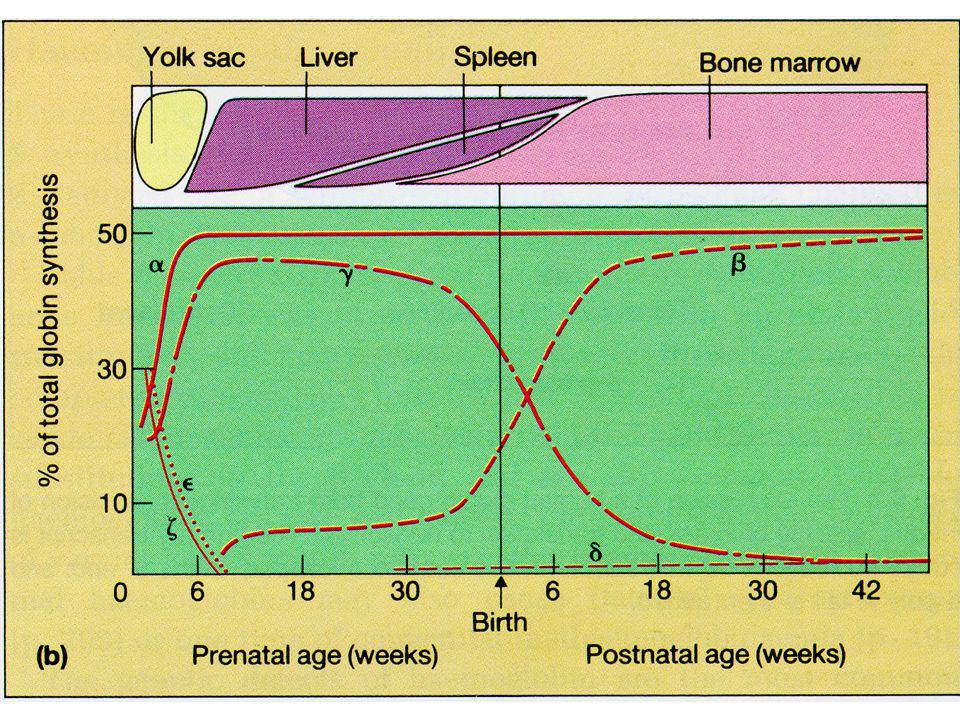
The Globin Genes

8
Hemoglobin Type Name Components Adult A 22 A2 22 Fetal F 22 Embryonic Portland 22 Gower 1 22 Gower 2 22 Abnormal H 4 Bart’s 4

9
Hemoglobin Types Quantity Alpha Thal >95% HbH HbA2 (α2 δ2) <3.5%
HbA (α2 β2) >95% HbH HbA2 (α2 δ2) <3.5% - HbF (α2 γ2) <2.0% Hb Barts
 >95% HbH. HbA2 (α2 δ2) <3.5% - HbF (α2 γ2) <2.0% Hb Barts.")
10
Hemoglobinopathy An inherited mutation of the globin genes leading to a quantitative or qualitative abnormality of globin synthesis

11
Thalassemia - Defined A family of genetic anemias characterized by a reduced rate of production of 1 or more globin subunits of hemoglobin (Hb) Symptoms are caused by the deleterious effects of the normally produced subunits that are now in excess

12
Pathophysiology Excess alpha chains precipitate and form inclusion bodies that associate with the RBC cell membrane Cause membrane damage and shortened cell survival Large scale destruction of precursor cells in bone marrow Decreased B production causes increased δ production and an elevated A2 (α2δ2)
")
13
Types of B-globin mutations
B0 – No B-globin chains are produced B+ - some beta chains produced Decreased

14
Alpha thalassemia AA / AA Normal AA / A -
Mild microcytosis (Silent Carrier) AA / - - A - / A - Mild microcytosis (Trait or Carrier) (cis vs trans) A - / - - Hemoglobin H disease- clinically variable - - / - - Hydrops Fetalis (Alpha Thal Major)
 AA / - - A - / A - Mild microcytosis (Trait or Carrier) (cis vs trans) A - / - - Hemoglobin H disease- clinically variable. - - / - - Hydrops Fetalis (Alpha Thal Major)")
15
α- and β-thalassemia Alpha Thalassemia
Deletions of alpha- globin gene (s) Symptoms can begin in fetal life Complicated inheritance – 4 alpha genes Beta Thalassemia Nonsense, splice and frameshift mutations in beta-globin gene Symptoms begin in infancy/childhood Simple AR inheritance; genotype-phenotype correlation
 Symptoms can begin in fetal life. Complicated inheritance – 4 alpha genes. Beta Thalassemia. Nonsense, splice and frameshift mutations in beta-globin gene. Symptoms begin in infancy/childhood. Simple AR inheritance; genotype-phenotype correlation.")
16
Structural variant - Defined
Abnormal globin protein that is produced at a normal rate, with varying consequences Oxygen affinity, stability and function

17
Normal

18
Normal Thalassemia

19
Normal Structural Variant

20
Laboratory Investigation
CBC-MCV,MCH,RDW Tests Hemoglobin electrophoresis Cellulose acetate: Alkaline pH Citrate agar: Acid pH Capillary Electrophoresis HPLC IEF DNA

21
Preanalytical EDTA sample Age H/O Transfusion Area of Residence E/D/C

22
SCREENING ANTENATAL PROFILE HbA1c ANEMIA PROFILE PRE MARITAL
NEONATAL PROFILE

23
Manual System

24
Cellulose Acetate pH-8.6 In a alkaline solution ,Hb molecules have a net negative charge and move towards the anode

25
Cellulose Acetate Hb Electrophoresis
- A F A + C/E/O S/D/G NORMAL BTT

26
Cellulose Acetate Hb Electrophoresis
- A2/C S F A+ Normal Hb AS Hb SS

27
Cellulose Acetate Hb Electrophoresis
- A2/C/E S /D F A+ Normal Hb AS HB AD

28
Citrate Agar Electrophoresis
Citrate agar electrophoresis at an acid Ph provides ready separation of hemoglobins that migrate together on cellulose acetate S from D and G C from E and O

29
Citrate Agar Hb Electrophoresis
C S A F _ Normal

30
Citrate Agar Hb Electrophoresis
C S A F _ Normal Sickle trait

31
HPLC Separation based on interaction between Stationary Phase & Mobile Phase Stationary Phase is Analytical Cartridge; Mobile Phase is Buffer Compounds are separated to target analytes according to physical properties: - size, shape, charge, hydrophobicity & affinity for other molecules Bound analytes elute off the stationary phase by manipulating the mobile phase

32
Contd… Charged particles (matrix) bind reversibly to sample molecules (proteins, etc.) Desorption is then brought about by increasing the salt concentration or by altering the pH of the mobile phase

33
Automated system;precalibrated column and gradient
HPLC Automated system;precalibrated column and gradient

34
Stationary Phase: Cation Exchange Cartridge
Direction of flow Detector Carboxyl groups attached to a resin base

35
Hemoglobin Introduction
Positively charged hemoglobin fragments in the hemolysate attach to the carboxyl groups at varying strengths.

36
Starting Gradient: Low Ionic Strength Buffer
The gradient starts with a low % of Buffer B (high % Buffer A) At this gradient, hemoglobin fragments with an ionic strength lower than the buffer gradient, such as A and F, are displaced from the cartridge and pass into the detector
 At this gradient, hemoglobin fragments with an ionic strength lower than the buffer gradient, such as A and F, are displaced from the cartridge and pass into the detector.")
37
Ending Gradient: High Ionic Strength Buffer
As the % of the High Ionic Strength Buffer B increases, the more hemoglobin fragments will be displaced Once the gradient is 100% Buffer B all remaining hemoglobin fragments, including any variant hemoglobins such as S, D and C, will be removed

38
CHROMATOGRAMS Peak Output Time Area RT

40
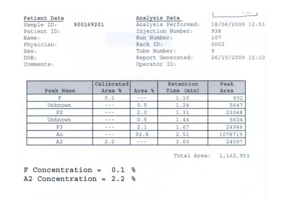
Normal HPLC Graph HbA F A2

41
Hb C Hb H HbF HbS Hb J Hb Köln HbE Hb O HbD Hb Q

42
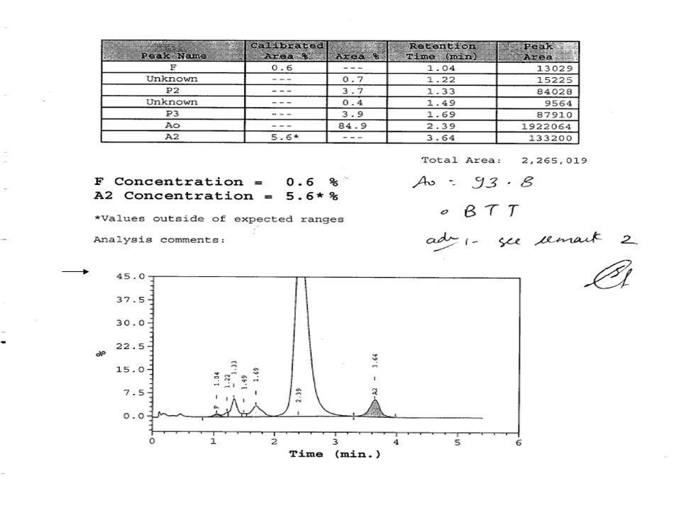
A2 A2 Suggests <3.5% Normal 3.5-8.0% BTT /(Megaloblastic anemia)
11-15% Hb Lepore 20-40% HbE 30-48% HbD

44
Hb Lepore

45
HbE

46
HbD

47
Hb EE/DD

48
HbF F Suggests <2% Normal <5% HPFH/Pregnancy/ Aplastic anemia
2-15% HPFH BTT Sickle 10-15% Delta Beta Thalassemia >50% Beta Thal Major HPFH (homozygous) Delta Beta Thalassemia (homo)
 Delta Beta Thalassemia (homo)")
49
Hb F 1.Delta beta Thal Trait 2.HPFH+IDA 1.Iron Studies 2.DNA Analysis

50
HPLC GRAPH OF CHILD Hb F= 100% Hb A2= 0% HbA0 =0% CBC: Hb = 6.8 g/dl
MCV = 67.3 Fl MCH = 21.3 pg RDW = 24.7%

51
HPLC GRAPH MOTHER Hb F = 7.0% HbA2 = 2.6% HbA0 = 90.4% CBC: Hb = 11.9
MCV = 64.7 MCH = 21.1 Impression: S/o Delta beta thal trait

52
HPLC GRAPH FATHER Impression S/O Delta beta thal trait HbF = 8.0%
HbA2= 2.8% HbA0 = 89.2% CBC: Hb = 13.4 MCV = 68.8 MCH = 22.4 Impression S/O Delta beta thal trait

53
HbF

54
Low A2 Iron Deficiency Anemia Alpha Thalassemia trait HbH Disease
Delta Thalassemia Delta –Beta Thalassemia

55
Peak before 1 min Sample Integrity Icteric sample HbH

56
HbH Age-8 yrs Hb-7.8g/dl MCV-61 fl MCH-17.8pg

57
HbH

58
Extended Retic stain HbH inclusions

59
Capillary Electrophoresis
Advantages HbH HbE vs HbD Disadvantages Preanalyticals (Cap piercing model) Interpretation
 Interpretation.")
60
Limitations…. Silent carriers Alpha Thalassemia
DNA analysis-gold standard

61
DNA analysis Silent carriers Prenatal period 5 common mutations

62
Conclusion….. Clinical Suspicion CBC PBS Reticulocyte count
Specific tests Electrophoresis HPLC Capillary Electrophoresis DNA analysis-Pre natal/gold standard

63
Questions ?

Similar presentations


 Korean J Hematol 46: 41-44 Microcytic.>")


 Hb is found in RBCs its main function is to transport O2 to tissues. Structure: 2 parts : heme + globin Globin: four globin chains (2 α.>")


 is composed of four protein chains, two α and two β globin chains arranged into.>")
 Units of energy Calorie.>")
