Download presentation
Presentation is loading. Please wait.

1
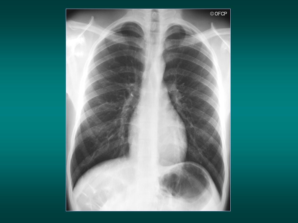
NORMAL CHEST X RAY

2
How to obtain a good quality chest radiography (1)
3 aspects are very important for good quality: The penetrating power of the x-ray beam (adjustment of x-ray tube voltage ) The x-ray tube current (milliampere) The exposure time adjustment
 The x-ray tube current (milliampere) The exposure time adjustment.")
3
How to obtain a good quality chest radiography (2)
The adjustment of the x-ray tube voltage controls the contrast: the difference of density levels of the different organs and tissues in the thorax The x-ray tube current and the exposure time controls the intensity of x-ray beams

4
How to obtain a good quality chest radiography (3)
Adjustment of voltage High voltage: a range of 100 /120 kV: optimal contrast between lungs and bones, and good visualisation of mediastinum and vessels Exposure time <0.05 seconds: decrease of motion artefact caused by the beating of the heart or respiratory movement

5
How to obtain a good quality chest radiography (4)
A long distance between the tube focus and the film improves the image clarity and decreases the geometric blur

6
How to obtain a good quality chest radiography (5)
Other criteria Quality of the x-ray grid: the flat metallic plate with very narrow lead trips close to the film: increase in the image clarity and reduction of the scattered radiation from the patient Good quality electrical power supply Efficient and frequent maintenance of x-ray equipment Quality of films and good conditions of storage Good screen-film system Good techniques for x-ray film processing (developing, rinsing, fixing, washing and drying procedure). If possible automatic film processor.
. If possible. automatic film processor.")
7
What about digital X ray system?(1) (Direct Digitalised System)
composed with: Electronic flat-panel X ray detector High resolution grayscale diagnostic display High performance computer

8
What about digital X ray system ?(2)
Avantages: -feasible imaging quality adjusted by computer processing -easy and quick image processing -X-ray film and its processing procedures in dark room no more needed. -Lower dose radiation - imaging transmission and intepretation via internet to referent radiologist is easily possible Disadvantages: - Costly initial investment (61000 to $) - Significant trainning in digital technology needed for the radiological technicians and costly running maintenance
 - Significant trainning in digital technology needed for the radiological technicians and costly running maintenance.")
9
Possible compromise with computed radiography (CR system)
digital radiography using a Photo‐stimulable Phosphor plate (PSP, also called Imaging Plate, IP) enclosed in a cassette as a detector, instead of a film‐screen. The IP is then introduced into a CR Reader which reads the film and converts the recorded signal into a digital grey scale image Same avantages than digitalised system Lower cost than DR system, because can be used with the existing X ray equipment. Only a CR reader and CR cassette reader (with Imaging Plates) need to be purchased..
 enclosed in a cassette as a detector, instead of a film‐screen. The IP is then introduced into a CR Reader which reads the film and converts the recorded signal into a digital grey scale image. Same avantages than digitalised system. Lower cost than DR system, because can be used with the existing X ray equipment. Only a CR reader and CR cassette reader (with Imaging Plates) need to be purchased..")
10
Basic radiographic views:
Postero-anterior view Lateral view Additional/supplementary view: radiography in expiration radiography in supine and lateral position Back view (antero-posterior view) Opacification of the oesaphagus
 Opacification of the oesaphagus.")
11
The thorax is composed of:
Bone (vertebrae, ribs, scapula…). The main component is calcium, which absorbs the x-ray considerably: the bone image is very opaque (white on the radiography) Blood and soft tissue (heart, mediastinum, vessels). The absorption of x-rays is less complete than bones: the image is less opaque (light grey) Fat tissue. the absorption of x-rays is lower: the image is dark grey. Air (in lungs) which does not absorb the x-ray at all. The image of the lungs is black
. The main component is calcium, which absorbs the x-ray considerably: the bone image is very opaque (white on the radiography) Blood and soft tissue (heart, mediastinum, vessels). The absorption of x-rays is less complete than bones: the image is less opaque (light grey) Fat tissue. the absorption of x-rays is lower: the image is dark grey. Air (in lungs) which does not absorb the x-ray at all. The image of the lungs is black.")
12
Picture of 4 different solutions on a chest x-ray film
calcium water oil Air

13
calcium Fluid / soft tissu air Fat

14
Dosage of x-rays type of investigation Equivalent of chest x-ray
Equivalent of natural radiation chest x-ray 1 3 days TDM 1 month-1year NMR: no radiation

15
Chest x-ray: criteria for quality
Deep inspiration Adequate contrast / density Good position of the patient X-ray beam in postero-anterior view (the patient is standing)
")
17
Same patient with deep inspiration Poor inspiration
False opacity of the inferior lobes Adequate inspiration if you can count 9 posterior or 6 anterior parts of ribs over the diaphragm

18
PA is the correct view false cardiomegaly AP versus PA view
clavicles are high and horizontal The x-ray beam is antero posterior Same patient with correct postero-anterior x-ray beam incidence PA is the correct view The technician should specify the position of the patient if AP view

19
The heart outline is bigger on D2
( bird’s-eye view of the patient)
")
20
Correct standing or sitting position for chest radiography

21
Standing or sitting position not always easy to obtain…

22
If the patient is in supine position (too ill to stand up), the cardiac outline and mediastinum is enlarged. The scapula may be on the lung field. The chest x-ray has poor quality for analysis

23
The technician should specify the patient position if supine
Standing patient with postero-anterior X ray beam Patient in supine position The technician should specify the patient position if supine

24
Dark film Lighy film no detail visible in the lung area
Under penetration No detail visible in the mediastinum area.

25
Correct density and good contrast:
- Pulmonary vessels visible in the lungs, behind the diaphragm and behind the heart Para-aortic line visible - Vertebra visible behind the mediastinum

26
Conditions for adequate contrast / density
Correct x-ray factor (Kv, Mas, exposure time) Good conditions of developing and good quality of processing chemicals Correct temperature of developer correct quality of film In case of digitalised or computerised system, imaging quality is adjusted by computer processing and the 3 last conditions are no more needed.
 Good conditions of developing and good quality of processing chemicals. Correct temperature of developer. correct quality of film. In case of digitalised or computerised system, imaging quality is adjusted by computer processing and the 3 last conditions are no more needed.")
27
Exact front view : the vertical line connecting the spinous process of thoracic vertebrae is in the middle of the two sterno-claviculars joints.

28
exact PA view left anterior oblique position

29
Front View l a o r a o D1 D2 D3 D3> D1>D2

30
Chest x-ray: to ensure top quality
deep inspiration adequate contrast / density correct position of the patient (exact front view) x-ray beam in postero anterior view (the patient is standing)
 x-ray beam in postero anterior view (the patient is standing)")
31
Process for analysis of the chest radiography: the check list
Verification of name and date Clinical history and findings Verification of the factors for good quality Assess -thoracic wall and thoracic skeleton - mediastinum - each lung field, one after the other Do not skip any item in the checklist !

33
Normal chest radiography
and some pitfalls… ( trouble-shooting)
")
34
Thoracic wall And skeleton

35
Sternocleidomastoid muscle
Thoracic wall Sus and retro clavicular field External side of Sternocleidomastoid muscle

36
thoracic wall 1 3 The clavicles are projected on the level of the 3rd or 4th posterior part of ribs 4

37
Cervical ribs: minor malformation

38
Be aware of foreign body or artefacts
on the chest x-ray trap picture: opacity of the superior part of right lung due to a hair braid

39
Breast implant

40
The retro-clavicular fields are always difficult to analyse, because of bone superposition:
- Clavicles - Anterior part of first rib - Posterior part of third and fourth rib, - Sterno-clavicular joint

41
There are 2 ways to correctly analyse the retro-clavicular fields:
Always compare right and left Ask for a chest x-ray with the patient’s back against the film (AP view, lordotic position)
")
42
Always compare left and right
Right TB infiltrate

43
Patient with fever, cough,
AFB sputum positive…

44
You have no CT scan. So use your eyes and compare right and left!
If any doubt, request AP/ lordotic chest X ray

45
Normal AP chest x-ray, back close to the film Normal chest x-ray , front close to the film

46
Chest x-ray, back close to the film And clavicles out of the field
by raising hands Chest x-ray, front close to the film

47
Chest x-ray back close to the film

48
physiological blur of the inferior side of the ribs
Thoracic wall physiological blur of the inferior side of the ribs Rib view section

49
You must always « read » a chest x-ray with methodology:
Example: for the chest wall, you must look at every rib, one after the other (6th and 9th ribs missing on this CXR)
")
50
Chest wall Top of the axillar hole Big pectoral muscle

51
thoracic wall Scapula

52
What is wrong with this chest x-ray?
Congenital clavicles agenesy

54
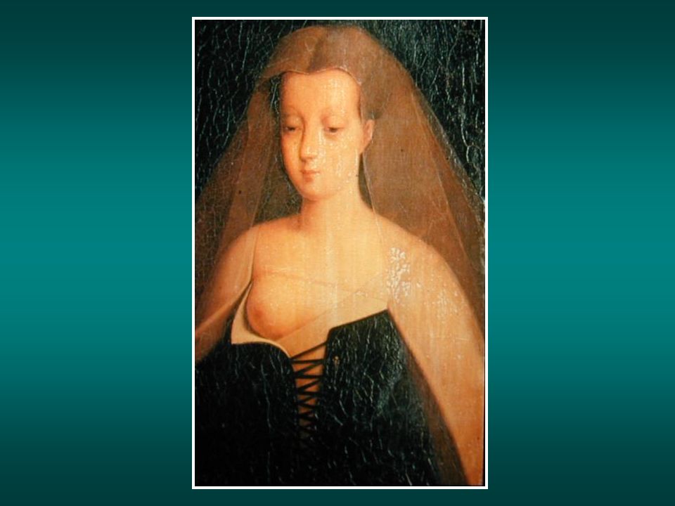
Thoracic wall Breast silhouette

55
Be careful with false opacities in the inferior lobes, consequences
of breast superposition.

56
Chest x-ray. Before and after right mastectomy

57
The right side is usually higher than the left side (3cm )
Thoracic wall Diaphragm The right side is usually higher than the left side (3cm )
")
59
Component elements of Mediatinum and hilus

60
AO AO PA LA RV RA RV LV LV

61
Front view Lateral view
AO SVC AO PA PA LV RA LA RV LV Front view Lateral view SVC: superor vena cava AO : aortic vessel PA: pulmonary artery RA:Right ventricle LV left ventricle LA: left auricle RV: right venticle LA RA LV RV

62
Left pulmonary artery Right pulmonary artery

63
The pulmonary vena are not physiologiccally visible
RSPV LPSV L A RIPV L V The pulmonary vena are not physiologiccally visible

64
Mediastinum lines X ray beam crossing thorax and mediastinum meets in some places pleural thickness, producing images on the CXR like lines defining mediastinum lines…

65
Too complicated And not so usefull… Mediastinum lines
1. Sub clavicle arterial line 2. Posterior mediastinum line 3. Brachio cepahalic vena line 4. para-azygos line 5. Anterior mediastinum line 6. Descending aortic line 7. Right and left paravertébral line 8. Inferior veina cava line 9. para-œsophageal line 10. para-trachéal line Too complicated And not so usefull… 6

66
Three of them are really important.
Right paratracheal line Aorto pulmonary line Para-aortic line Three of them are really important.

67
Mediastinum enlargment due to fat tissu

68
Be aware of false enlargment of mediastinum if obesity, poor inspiration, oblique view or supine position

69
Trap: false mediastinum enlargment in the case of this older woman with cyphoscoliosis, in supine position

71
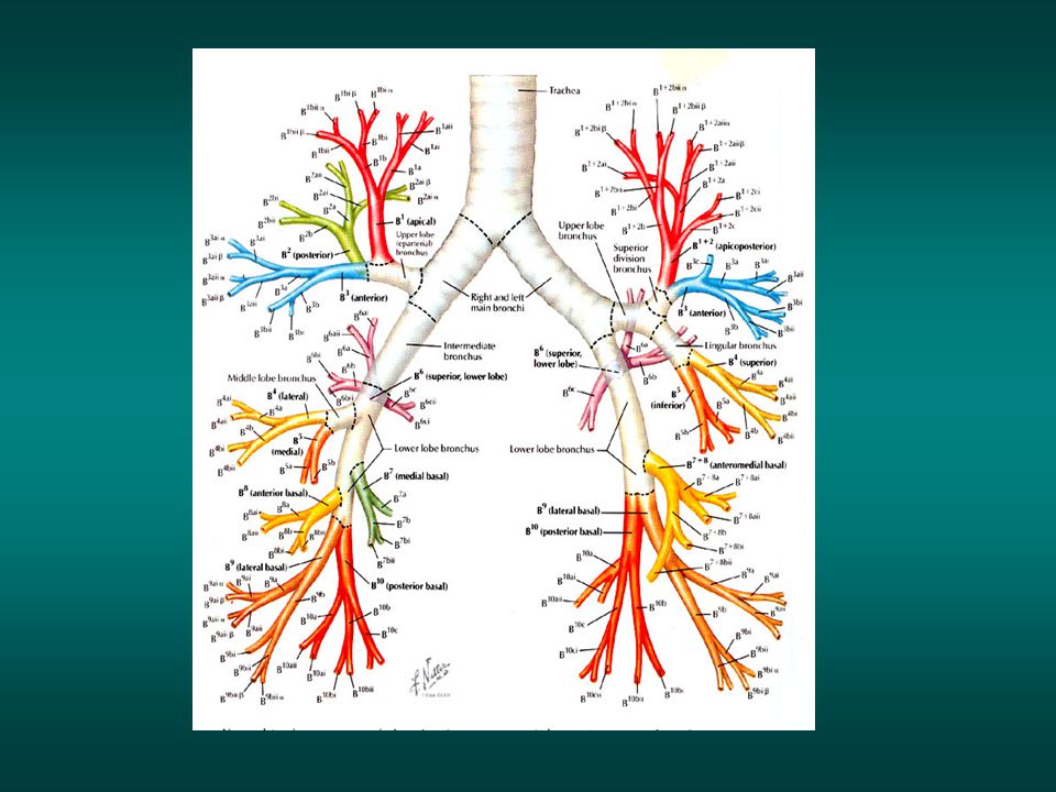
Component elements of lungs

72
On a normal chest x-ray, bronchi are not visible
(opacification with iodin hydrosoluble solution)
")
73
…but pulmonary arteries are visible.

76
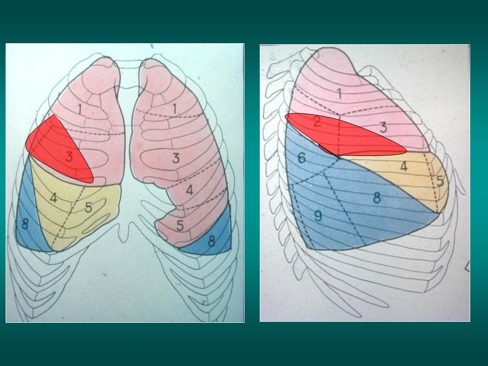
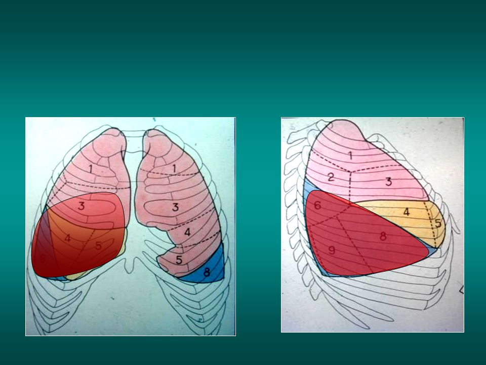
Small fissura Big fissura Right view

77
Left fissura Left view

78
Right superior lobe pneumonia
minor fissura Right superior lobe pneumonia minor fissura Large oblique fissura posterior part

80
Right inferior pneumonia
Large oblique fissura

82
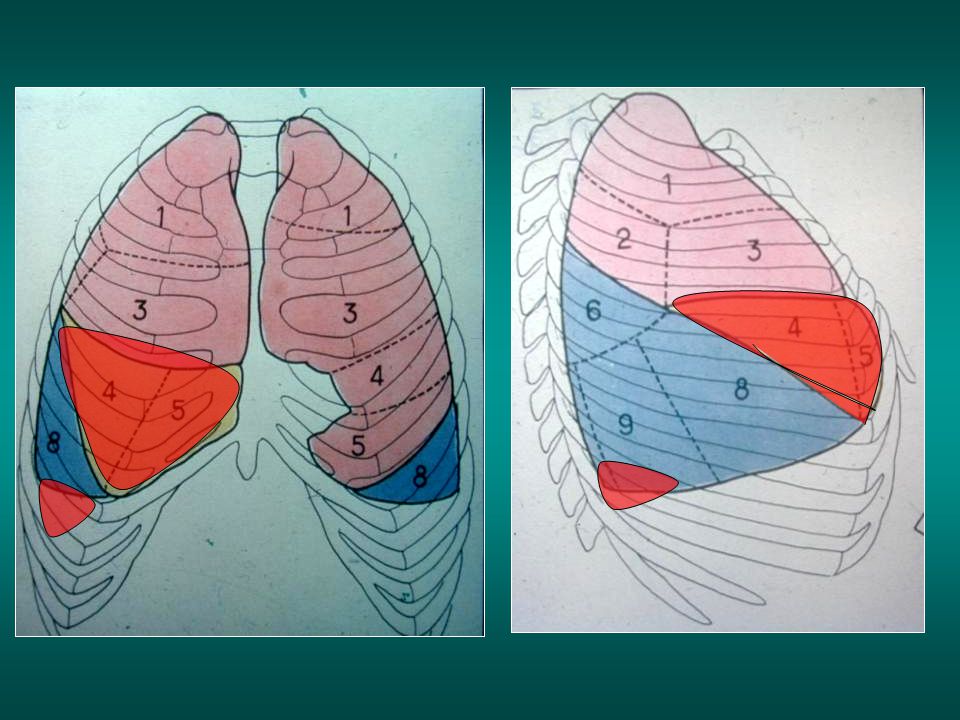
Middle lobe pneumonia Large fissura minor fissura
Small pleural effusion

84
External segment of middle lobe pneumonia

85
External segment of middle lobe pneumonia

86
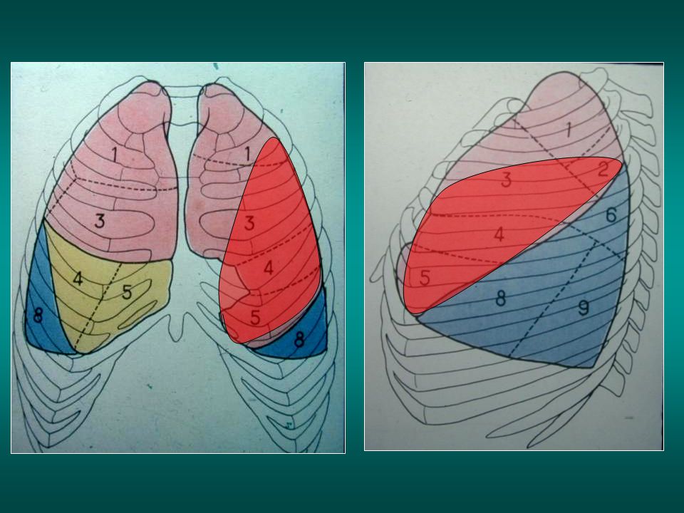
Left superior lobe pneumonia

88
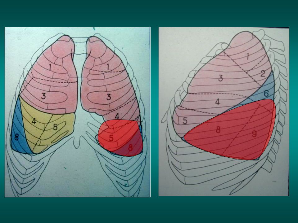
Left inferior pneumonia
Left scissura

91
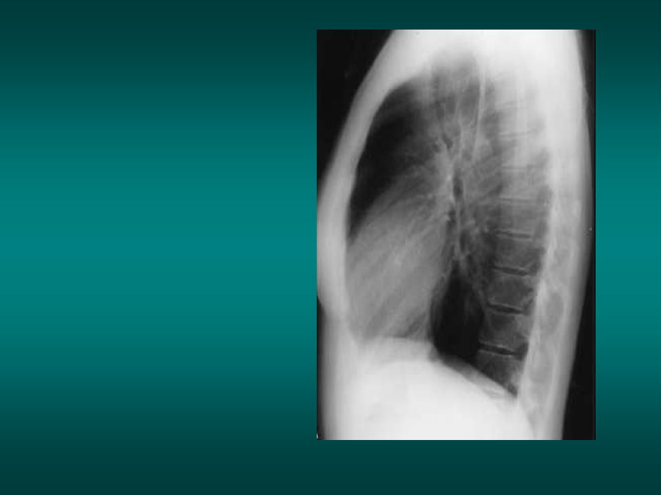
Normal lateral view

92
Lateral view

93
AO PA LA RV LV

94
Front view Lateral view
AO SVC AO PA PA LV RA LA RV LV Front view Lateral view LA RA LV RV

95
Heart and Mediastinum vessels
Superior vena cava Heart and Mediastinum vessels Ascending Aorta Pulmonary arteria Right ventricle Descending aorta

96
Heart and Mediastinum vessels Left ventricle Inferior vena cava

97
Mediastinum vessels Aortic arch

98
mediastinum vessels Descending aorta

99
Right pulmonary artery
Mediastinum vessels Right pulmonary artery Left pulmonary artery

100
normal CXR Lateral view is very useful for diagnosis of mediastinal adenopathies

101
trachea 20 mm

102
Right superior lobe bronchus Left superior lobe bronchus

104
The «clear spaces» Retro sternal clear space Retro cardiac clear space

105
The «clear spaces» Retro tracheal space

106
enlargment of the clear spaces: Emphysema

107
Emphysema Normal lateral view

108
The retro sternal space is filled: thymoma

109
The retro sternal space is filled: thymoma. Normal view on the right

110
Diaphragm Right diaphragm Left diaphragm

111
Summary normal CXR

112
Normal frontal chest view Aortic arch (AA) Descending aorta (DA)
Proximal left pulmonary artery (PLPA) Left interlobar pulmonary artery (LPA) Aortopulmonary window (AW) Left main bronchus (LB) Left atrial appendage (LAA) Left ventricle (LV) Superior vena cava (SVC) Right paratracheal stripe (RPS) Azygos vein (AV) Right interlobar pulmonary artery (RIPA) Right atrium (RA) Trachea (T) Right diaphragm (RD) Left diaphragm (LD) T T SVC SVC A A RPTS A W AV LPA RIPA LB DA RA RD LV LD
 Left interlobar pulmonary. artery (LPA) Aortopulmonary window (AW) Left main bronchus (LB) Left atrial appendage (LAA) Left ventricle (LV) Superior vena cava (SVC) Right paratracheal stripe. (RPS) Azygos vein (AV) Right interlobar pulmonary. artery (RIPA) Right atrium (RA) Trachea (T) Right diaphragm (RD) Left diaphragm (LD) T. T. SVC. SVC. A A. RPTS. A W. AV. LPA. RIPA. LB. DA. RA. RD. LV. LD.")
113
Normal lateral chest view
Posteriorly: vertebral bodies (V) and intervertebral disc spaces (*) Anteriorly: retrosternal clear space (RSCS) Trachea (T) Orifice of Right Upper Lobe bronchus (RUL) appears as circular lucency projecting over the continuation of the tracheal air column Left pulmonary artery (LPA) Right pulmonary artery (RPA) Left auricle (LA) Left ventricle (LV) Right ventricle (RV) Aortic arch (AA) Postero costo phrenic angle (PCPA) Right diphragm (RD) Left diaphragm (LD) Inferior vena cava (IVC) Scapula (Sc) Retro cardiac clear space (RCCS) T Sc AA RSCS RPA LPA RUL RV LA V RCCS * LV IVC LD RD PCPA
 and intervertebral disc spaces (*) Anteriorly: retrosternal clear space (RSCS) Trachea (T) Orifice of Right Upper Lobe bronchus (RUL) appears as circular lucency projecting over the continuation of the tracheal air column. Left pulmonary artery (LPA) Right pulmonary artery (RPA) Left auricle (LA) Left ventricle (LV) Right ventricle (RV) Aortic arch (AA) Postero costo phrenic angle (PCPA) Right diphragm (RD) Left diaphragm (LD) Inferior vena cava (IVC) Scapula (Sc) Retro cardiac clear space (RCCS) T. Sc. AA. RSCS. RPA. LPA. RUL. RV. LA. V. RCCS. * LV. IVC. LD. RD. PCPA.")
Similar presentations