Download presentation
Presentation is loading. Please wait.

1
FUNGI OF MEDICAL IMPORTANCE.
There are many way to classify fungi, they can be classified according to their cellular organization and the way they reproduce, also by the site in which can cause disease. Each type has a different site of the body to infect and a different pattern of disease characteristics.

2
Basic morphological and clinical classification of the medically important Fungi

3
Superficial mycoses: These are superficial cosmetic fungal infections of the Hair, dead skin and lipids secretions. Fungi which cause this kind of infection are living as saprophytes, They don’t provoke any immune response from the host. No living tissue is invaded. Essentially no pathological changes are elicited. These infections are often so harmless that patients are often unaware of their condition. The important superficial mycosis includes: Ringworm (dermatophytosis), caused by Microsporum species, Trichophyton species, Epidermophyton Floccosum. Pityriasis versicolor (tinea versicolor). Piedra (trichosporosis) caused by Piedraia species
, caused by Microsporum. species, Trichophyton species, Epidermophyton Floccosum. Pityriasis versicolor (tinea versicolor). Piedra (trichosporosis) caused by Piedraia species.")
4
Dermatophytosis: Ringworm infections are the commonest of all mycoses. Ringworm is often referred to clinically as Tinea. Dermatophytosis: =Tinea = Ringworm infection. Infection of the skin, hair or nails caused by a group of keratinophilic fungi, called dermatophytes. Dermatophytes : are a group of fungi that infect the superficial keratinized portion of the body- skin, hair, nails without spread into the deeper skin Dermatophytosis is caused by 17 species of microsporum, 22species of Trichophyton, or by Epidermophyton Floccosum. Microsporum: Hair, skin Epidermophyton: Skin, nail Trichophyton: Hair, skin, nail

5
Ringworm fungi infect only the keratinized layers of the skin, hair and nails due to their ability to obtain nutrients from keratinized material. They are usually restricted to the nonliving layer of the epidermis because of their inability to penetrate viable tissue of an immunocompetent host. Acid proteinases, elastase, keratinases, and other proteinases reportedly act as virulence factors. The organisms colonize the keratin tissues and inflammation could be caused by host response to metabolic by-products. The spores settle on the skin, germinate and form a mass of branching hyphae, which grow out radially to produce circular lesions.

6
Dermatophytes classification:
Classified into three groups depending on their usual habitat: Anthropophilic dermatophytes are restricted to human hosts and produce a mild, chronic inflammation. Zoophilic organisms are found primarily in animals and cause marked inflammatory reactions in humans who have contact with infected cats, dogs, cattle, horses, birds, or other animals. This is followed by a rapid termination of the infection. Geophilic species are usually recovered from the soil but occasionally infect humans and animals. They cause a marked inflammatory reaction, which limits the spread of the infection and may lead to a spontaneous cure but may also leave scars

7
Dermatophytosis clinical classification:
Infection is named according to the anatomic location involved: Ringworm Favus Onychomycosis

8
Ring worm infections Dermatophytosis clinical manifestation Skin: Circular dry lesions Slightly raised red scaly margins Surrounded by red itchy skin Metabolites provoke inflammation Hair: typical lesions → scarring Nail: thickened, deformed, friable, discoloured Hair invasion: Endothrix Ectothrix Favus

9
Favus ( Endothrix Favic hair infection):
A special type of endothix hiar infection is caused by T. schoenleinii. The hyphae do not break up into erthrospores, but eventually die leaving characteristic air spaces. The hair doesn’t break, but lost completely and permanently due to degeneration of follicle Sharp delimited bald spots Adjacent skin is invaded, crusty and scarred

10
3- Onychomycosis Chronic infection of nail and nail-bed Most common in toenails Distal part gradually breaks and crumbles Main causative agents: T. rubrum T. interdigitale

11
Dermatophytosis transmission:
Dermatophytes are transmitted by: Direct contact with infected host (human or animal) or By direct or indirect contact with infected skin or hair in clothing, combs, hair brushes, caps, furniture, bed linens, towels, and hotel rugs. Depending on the species the organism may be viable in the environment for up to 15 months. There is an increased susceptibility to infection when there is a preexisting injury to the skin such as scars, burns, excessive temperature and humidity.
 or. By direct or indirect contact with infected skin or hair in clothing, combs, hair brushes, caps, furniture, bed linens, towels, and hotel rugs. Depending on the species the organism may be viable in the environment for up to 15 months. There is an increased susceptibility to infection when there is a preexisting injury to the skin such as scars, burns, excessive temperature and humidity.")
12
Dermatophytosis diagnosis:
Clinical diagnosis Laboratory diagnosis. Physiological tests

13
Dermatophytosis clinical diagnosis
Appearance Clinical information such as the appearance of the lesion, site, geographic location, travel history, animal contacts and race is also important, especially in identifying rare non-sporulating species 2- Dermatophytosis laboratory diagnosis: Ringworm fungi are monomorphic MOLDS, the laboratory diagnosis is best made by direct microscopy. Culture is required to identify the infecting species.

14
Collection of the specimens:
Clean the affected area with 70% v/v ethanol. Collect skin scales, crust, pieces of nails or hair on a clean piece of paper. Skin scales: collect by scraping the surface of the margin of the lesion using sterile blade. Crusts: collect by removing part of the crust nearest to healthy skin using sterile scissors and tweezers. Nail pieces: collect by taking snipping of the infected part of the nail using sterile scissors. Hair: collect by Completely removing affected of fluorescent dull broken hairs from the margin of the lesion using sterile tweezers, massage brush Or adhesive tape

15
Use cotton swab for: Moist skin area Mucus membranes External ears After collecting the specimens, give name and number of the patient, date of collection, and any other data. Transfer it to the mycology lab. Note: ringworm specimens are best transported in paper than in screw-cap container, to reduce the humidity and the multiplication of bacteria. Spore resist drying and remaining viable for several weeks when stored in paper.

16
Direct microscopic This is the recommended method to diagnose ringworm (10-20% of specimens were positive in direct microscopy, but negative on culture). The material from skin, nails, etc. is first softened and cleared with a strong alkali such as 200g/l KOH, which will digest the keratin surrounding the fungi so that the hyphae and spores can be seen. Hairs clean within 10 min, skin scales and crusts usually take min, and nails may take several hours to clear. Clearing can be hastened by gently heating. As soon as the specimen has cleared examine it microscopically using the 10x and 40x objectives. Fungi are usually larger than bacteria.
. The material from skin, nails, etc. is first softened and cleared with a strong alkali such as 200g/l KOH, which will digest the keratin surrounding the fungi so that the hyphae and spores can be seen. Hairs clean within 10 min, skin scales and crusts usually take min, and nails may take several hours to clear. Clearing can be hastened by gently heating. As soon as the specimen has cleared examine it microscopically using the 10x and 40x objectives. Fungi are usually larger than bacteria.")
17
Appearance of hair in KOH preparation:
Look for arthrospores and hyphae, and note whether infection is on the outside of the hair or within it. When it is in the outside, it is referred to as ectothrix. The arhtrospores may be arranged in chain or in masses around the hair. When the infection is within the hair, it is referred to as endothrix. Characteristic hyphae can be seen spread among the epithelial cells. Trichophyton tonsurans, the causative agent of tinea capitis (scalp infection) can be seen as solidly packed arthrospores within the broken hairshafts scraped from the plugged black dots of the scalp.
 can be seen as solidly packed arthrospores within the broken hairshafts scraped from. the plugged black dots of the scalp.")
18
Culture: Fungal culture medium is used for positive identification of the species. It is usually done using culture which encourage hyphae growth and spore formation, such a medium should inhibit the bacterial and saprophytic fungi growth. Usually fungal growth is noted in 5 to 14 days. Culture characteristics such as surface texture and pigmentation are variable so they are the least reliable criteria for identification. A special media called Dermatophyte Test Medium (DTM) has been formulated to grow and identify dermatophytes. Without having to look at the colony, the hyphae, or macroconidia - one can identify the dermatophyte by a simple color test.
 has been formulated to grow and identify dermatophytes. Without having to look at the colony, the hyphae, or macroconidia - one can identify the dermatophyte by a simple color test.")
19
The specimen (scraping from skin, nail, or hair) is embedded in the DTM culture medium. It is incubated at room temperature for 10 to 14 days. If the fungus is a dermatophyte, the medium will turn bright red. If the fungus is not a dermatophyte, no color change will be noted . If kept beyond 14 days, false positive can result even with non-dermatophytes. Specimen from the DTM can be sent for species identification if desired There are many other medium that can be used for the identification of the dermatophyte as Malt Extract Agar which contains cycloheximide and antibiotic such as chloramphenicol. Another media which could be used is Sabouraud dextrose agar. Plate culture is recommended more than slope, because a heavier inoculum can be cultured and more air is available which helps fungus to grow(dermatophytes are aerobic). To prevent drying of the medium, it should be kept in sealed plastic bags.
. To prevent drying of the medium, it should be kept in sealed plastic bags.")
20
Skin scales, crusts, pieces of nail:
Using a sterile blade or scissors, cut the specimens into pieces as small as possible. Using sterile tweezers, inoculate the small pieces (a few mm apart), on the surface of the plate of malt agar. Inoculate at room temperature (25-30 oC) for up to3 weeks, examining every few days for growth. Hairs: 1- Using a sterile scissor cut the hair into small pieces about 3-5 mm long. 2- Using sterile tweezers, inoculate the pieces of the hair in the surface of the plate of malt agar. 3-Inoculate at room temperature (25-30 oC) for up to3 weeks, examining every few days for growth.
, on the surface of the plate of malt agar. Inoculate at room temperature (25-30 oC) for up to3 weeks, examining every few days for growth. Hairs: 1- Using a sterile scissor cut the hair into small pieces about 3-5 mm long. 2- Using sterile tweezers, inoculate the pieces of the hair in the surface of the plate of malt agar. 3-Inoculate at room temperature (25-30 oC) for up to3 weeks, examining every few days for growth.")
21
Examination of the culture:
The following features are used in identification of ringworm fungi species: Hyphal characteristics such as spiral, pectinate, or antler hyphae. Absence or presence of spores. Morphology of spores. Microscopy from cultures: Mounting media or Cello tape (Scotch) method. Water or Lactophenol-cuttonblue.
 method. Water or Lactophenol-cuttonblue.")
22
Mounting media. Using a sterile loop, remove carefully a portion of the colony and transfer it to a drop of ethanol on a slide. The ethanol prevents the trapping of air bobbles. When ethanol evaporates, add a drop of lactophenol cotton blue, and cover it with a cover glass. Examine the preparation using the 10X and 40X objectives. Look for the presence of macroconidia, microconidia, chlamydospores, and hyphae which appear spiral, pectinate, or antler-like, reflexive, or markedly distorted.

23
2- Cello tape (Scotch) method:
The usual wet mount method is a helpful method in the diagnosis of fungi, it helps if characteristic spores are found, but it is not suitable to identify the characteristic spores arrangement. The most helpful procedure is the use of adhesive transparent tape (scotch tape) method. The adhesive side of the tape is gently touched to the colony and then is adhered to a slide in which a drop of lactophenol cotton blue has been placed. This procedure allow you to observe the spores and supporting structures arranged somewhat as they were when part of the colony.
 method. The adhesive side of the tape is gently touched to the colony and then is adhered to a slide in which a drop of lactophenol cotton blue has been placed. This procedure allow you to observe the spores and supporting structures arranged somewhat as they were when part of the colony.")
25
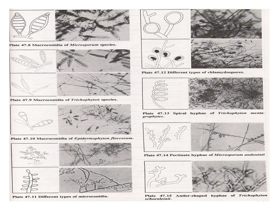
Macroconidium and microconidium
Macroconidia: These are large spores which vary in appearance with the different species as follows: 1- Microsporum: Macroconidia are spindle-shaped with slightly tapering ends, symmetrical, rough-surfaced and thick-walled. They may be very large with up to 15 septa.

26
2- Trichophyton: macroconidia vary in morphology
2- Trichophyton: macroconidia vary in morphology. They may appear cylindrical or club-shaped. They are smooth-surfaced, thin-walled, and may show elongated ends, with up to 8 septa.

27
3- Epiderophyton: macroconidia are club-shaped, or oval, smooth-surfaced, with moderately thick-walls, and only few septa.

28
Microconidia: These are small, round, oval or elongated spores arising from the ends or sides of hyphae. They are often seen in clusters.

29
Chlamydospores: These are round spores of varying shape and size. .

30
Spiral hyphae: These are arising from the side or the ends of thickened hyphae.

31
Pectinate hyphae: These are thickened, distorted hyphae, with protrusions. Antler hyphae: These are short hyphae with branching ends, which look like stag antlers.

32
Reflexive hyphae: These are hyphae which bend in a backwards direction

33
Characteristics morphology
T. mentagrophytes “spiral hyphae” T. schoenleinii “antler-like hyphae” M. rivalieri “ pectinate hyphae” C. Physiological tests to differentiate between the species: In vitro hair perforation test Special amino acid and vitamin requirements Urea hydrolysis Growth on many different special media Temperature tolerance and enhancement

34
Serology: serological tests are not appropriate for the diagnosis of superficial fungal infections.

Similar presentations

>")


>")